9LAD | pdb_00009lad

Crystal structure of the complex between Neuronal Pentraxin 2 (NP2 PTX) and antibody fragment VHH N1


Experimental Data Snapshot

  • Method: X-RAY DIFFRACTION
  • Resolution: 2.02 Å
  • R-Value Free: 
    0.220 (Depositor), 0.228 (DCC) 
  • R-Value Work: 
    0.182 (Depositor), 0.189 (DCC) 

Starting Model: in silico
View more details

wwPDB Validation 3D Report Full Report

Validation slider image for 9LAD

This is version 1.1 of the entry. See complete history

Literature

Development of a VHH that inhibits the binding of neuronal pentraxin 2 to a postsynaptic glutamate receptor, AMPAR.

Yokoo, T.Nakakido, M.Matsuda, K.Caaveiro, J.M.M.Fernandez-Perez, J.Yuzaki, M.Tsumoto, K.

(2025) J Biological Chem 302: 110975-110975

  • DOI: https://doi.org/10.1016/j.jbc.2025.110975
  • Primary Citation Related Structures: 
    9LAD

  • PubMed Abstract: 

    Neurons connect to each other via synapses to form neural circuits. Recent research has shown that neuropsychiatric disorders and neurological disorders such as autism spectrum disorders and Alzheimer's disease (AD) are synaptic diseases caused by abnormality of synapses. Synaptic organizers are molecules responsible for synapse formation. Neuronal pentraxin 2 (NP2) is synaptic organizer and a secreted protein that is expressed mainly in the hippocampus and cerebellum, and it contributes to synaptic plasticity. NP2 forms clusters with its family proteins NP1 and NPR and binds to postsynaptic amino-3-hydroxy-5-methyl-4-isoxazolepropionic acid-type receptors (AMPARs). In recent years, research has revealed the disease relevance of NP2. For example, it can be a biomarker of AD, and its overexpression in the peripheral nervous system has been reported to cause chronic itch. However, the mechanism of NP2 function has not been well described at the molecular level. In this study, we developed a variable domain of heavy chain of heavy chain antibody (VHH) against NP2 to elucidate its molecular mechanism of action and to regulate its function of NP2. The obtained VHH N1 showed high specificity and affinity to NP2, and its binding mechanism was elucidated by X-ray crystallography. Furthermore, VHH N1 inhibited the binding of NP2 to AMPARs, and this inhibitory activity was confirmed in cells. These results provide useful insights into the molecular mechanism of NP2 function and highlight the potential application of VHH N1 as a detection agent for NP2 or as a therapeutic agent for chronic itch.


  • Organizational Affiliation
    • Department of Chemistry and Biotechnology, School of Engineering, The University of Tokyo, 7-3-1, Hongo, Bunkyo-ku, Tokyo, 113-8656, Japan.

Macromolecule Content 

  • Total Structure Weight: 38.56 kDa 
  • Atom Count: 2,702 
  • Modeled Residue Count: 326 
  • Deposited Residue Count: 345 
  • Unique protein chains: 2

Macromolecules

Find similar proteins by:|  3D Structure
Entity ID: 1
MoleculeChains  Sequence LengthOrganismDetailsImage
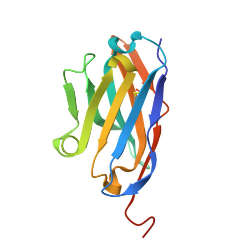
N1 VHHA [auth B]126Vicugna pacosMutation(s): 0 
Find similar proteins by:|  3D Structure
Entity ID: 2
MoleculeChains  Sequence LengthOrganismDetailsImage
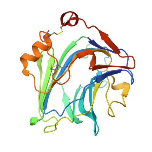
Neuronal pentraxin-2B [auth C]219Homo sapiensMutation(s): 0 
Gene Names: Nptx2Narp
UniProt
Find proteins for P97738 (Rattus norvegicus)
Explore P97738 
Go to UniProtKB:  P97738
Entity Groups
Sequence Clusters30% Identity50% Identity70% Identity90% Identity95% Identity100% Identity
UniProt GroupP97738
Sequence Annotations
Expand
Reference Sequence

Small Molecules

Ligands 4 Unique
IDChains Name / Formula / InChI Key2D Diagram3D Interactions
GOL

Query on GOL


Download:Ideal Coordinates CCD File
G [auth C]GLYCEROL
C3 H8 O3
PEDCQBHIVMGVHV-UHFFFAOYSA-N
AZI

Query on AZI


Download:Ideal Coordinates CCD File
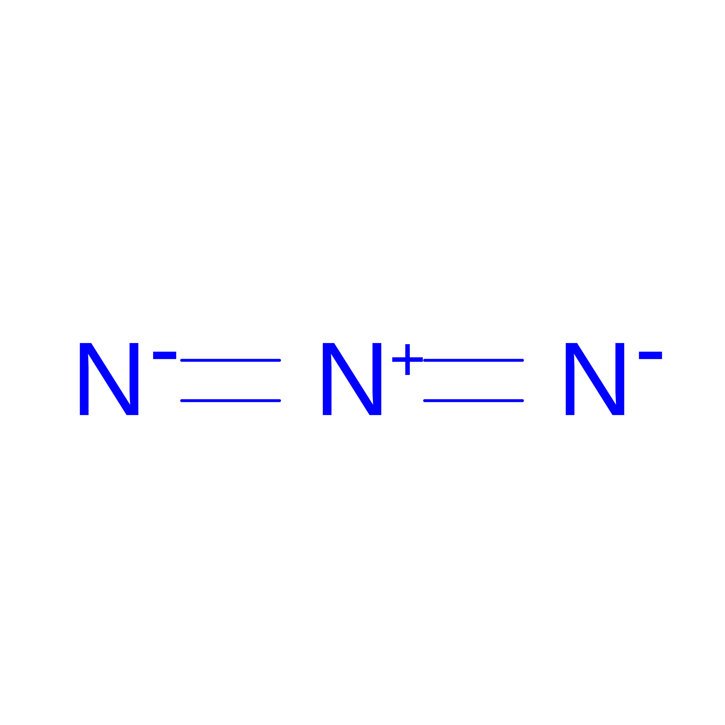
C [auth B]AZIDE ION
N3
IVRMZWNICZWHMI-UHFFFAOYSA-N
CA
(Subject of Investigation/LOI)

Query on CA


Download:Ideal Coordinates CCD File
D [auth C],
E [auth C]
CALCIUM ION
Ca
BHPQYMZQTOCNFJ-UHFFFAOYSA-N
CL

Query on CL


Download:Ideal Coordinates CCD File
F [auth C],
H [auth C]
CHLORIDE ION
Cl
VEXZGXHMUGYJMC-UHFFFAOYSA-M

Experimental Data & Validation

Experimental Data

  • Method: X-RAY DIFFRACTION
  • Resolution: 2.02 Å
  • R-Value Free:  0.220 (Depositor), 0.228 (DCC) 
  • R-Value Work:  0.182 (Depositor), 0.189 (DCC) 
Space Group: P 21 21 2
Unit Cell:
Length ( Å )Angle ( ˚ )
a = 93.574α = 90
b = 118.234β = 90
c = 39.464γ = 90
Software Package:
Software NamePurpose
REFMACrefinement
XDSdata reduction
Aimlessdata scaling
PHASERphasing

Structure Validation

View Full Validation Report


Entry History 

& Funding Information

Deposition Data


Funding OrganizationLocationGrant Number
Japan Society for the Promotion of Science (JSPS)Japan21H05090
Japan Society for the Promotion of Science (JSPS)Japan20H02531
Japan Society for the Promotion of Science (JSPS)Japan23KJ0516
Japan Agency for Medical Research and Development (AMED)Japan18ak0101100
Japan Agency for Medical Research and Development (AMED)JapanJP223fa627001
Japan Agency for Medical Research and Development (AMED)JapanJP223fa727002
Japan Agency for Medical Research and Development (AMED)JapanJP22ama121033
Japan Agency for Medical Research and Development (AMED)JapanJP24ama121031

Revision History  (Full details and data files)

  • Version 1.0: 2025-12-10
    Type: Initial release
  • Version 1.1: 2026-01-07
    Changes: Database references