9L8M | pdb_00009l8m

Structure of SID-1 in complex with dsRNA


Experimental Data Snapshot

  • Method: ELECTRON MICROSCOPY
  • Resolution: 2.90 Å
  • Aggregation State: PARTICLE 
  • Reconstruction Method: SINGLE PARTICLE 

wwPDB Validation 3D Report Full Report

Validation slider image for 9L8M

This is version 1.0 of the entry. See complete history

Literature

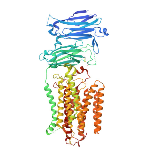
Structural insights into SID1-mediated dsRNA uptake: A self-organizing endocytic mechanism

Takai, A.Kumazaki, K.Awazu, T.Kambara, T.Murakoshi, S.Kato, T.Hiraizumi, M.Kise, Y.Kusakizako, T.Nishizawa, T.Okada, T.Nureki, O.

To be published.

Macromolecule Content 

  • Total Structure Weight: 96.24 kDa 
  • Atom Count: 5,652 
  • Modeled Residue Count: 653 
  • Deposited Residue Count: 783 
  • Unique protein chains: 1
  • Unique nucleic acid chains: 2

Macromolecules


Find similar proteins by:|  3D Structure
Entity ID: 1
MoleculeChains  Sequence LengthOrganismDetailsImage
Systemic RNA interference defective protein 1761Caenorhabditis elegansMutation(s): 0 
Gene Names: sid-1rde-7rsd-8C04F5.1
UniProt
Find proteins for Q9GZC8 (Caenorhabditis elegans)
Explore Q9GZC8 
Go to UniProtKB:  Q9GZC8
Entity Groups
Sequence Clusters30% Identity50% Identity70% Identity90% Identity95% Identity100% Identity
UniProt GroupQ9GZC8
Glycosylation
Glycosylation Sites: 3
Sequence Annotations
Expand
Reference Sequence
Find similar nucleic acids by:  Sequence
Entity ID: 2
MoleculeChains LengthOrganismImage
RNA (24-mer)B [auth C]11synthetic construct
Sequence Annotations
Expand
Reference Sequence
Find similar nucleic acids by:  Sequence
Entity ID: 3
MoleculeChains LengthOrganismImage
RNA (24-mer)C [auth D]11synthetic construct
Sequence Annotations
Expand
Reference Sequence

Oligosaccharides

Help  
Entity ID: 4
MoleculeChains Length2D Diagram GlycosylationD Interactions
2-acetamido-2-deoxy-beta-D-glucopyranose-(1-4)-2-acetamido-2-deoxy-beta-D-glucopyranoseD [auth B],
E
2N-Glycosylation
Glycosylation Resources
GlyTouCan: G42666HT
GlyCosmos: G42666HT
GlyGen: G42666HT

Small Molecules

Ligands 4 Unique
IDChains Name / Formula / InChI Key2D Diagram3D Interactions
LBN

Query on LBN


Download:Ideal Coordinates CCD File
I [auth A],
J [auth A]
1-palmitoyl-2-oleoyl-sn-glycero-3-phosphocholine
C42 H82 N O8 P
WTJKGGKOPKCXLL-VYOBOKEXSA-N
Y01

Query on Y01


Download:Ideal Coordinates CCD File
H [auth A]CHOLESTEROL HEMISUCCINATE
C31 H50 O4
WLNARFZDISHUGS-MIXBDBMTSA-N
NAG

Query on NAG


Download:Ideal Coordinates CCD File
F [auth A]2-acetamido-2-deoxy-beta-D-glucopyranose
C8 H15 N O6
OVRNDRQMDRJTHS-FMDGEEDCSA-N
ZN

Query on ZN


Download:Ideal Coordinates CCD File
G [auth A]ZINC ION
Zn
PTFCDOFLOPIGGS-UHFFFAOYSA-N

Experimental Data & Validation

Experimental Data

  • Method: ELECTRON MICROSCOPY
  • Resolution: 2.90 Å
  • Aggregation State: PARTICLE 
  • Reconstruction Method: SINGLE PARTICLE 
EM Software:
TaskSoftware PackageVersion
MODEL REFINEMENTServalcat0.4.88
RECONSTRUCTIONRELION3.1

Structure Validation

View Full Validation Report


Entry History 

& Funding Information

Deposition Data


Funding OrganizationLocationGrant Number
Japan Society for the Promotion of Science (JSPS)JapanJP21H05037
Japan Science and TechnologyJapanJPMJCR20E2
Japan Agency for Medical Research and Development (AMED)JapanJP19am01011115

Revision History  (Full details and data files)

  • Version 1.0: 2026-01-21
    Type: Initial release