


Crystal structure of Klebsiella pneumoniae Enoyl-Acyl Carrier Protein Reductase (FabI) in complex with Triclosan
Biswas, S., Patra, A., Kushwaha, G.S., Suar, M.To be published.
Experimental Data Snapshot
Starting Model: experimental
View more details
Entity ID: 1 | |||||
|---|---|---|---|---|---|
| Molecule | Chains | Sequence Length | Organism | Details | Image |
| Enoyl-[acyl-carrier-protein] reductase [NADH] | 265 | Klebsiella pneumoniae subsp. pneumoniae 12-3578 | Mutation(s): 0 Gene Names: fabI, E9161_16145, GLO21_010850, GLO21_17500, NCTC9504_03879 EC: 1.3.1.9 | 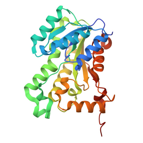 | |
UniProt | |||||
Find proteins for A0A1Y0Q1M7 (Klebsiella pneumoniae subsp. pneumoniae) Explore A0A1Y0Q1M7 Go to UniProtKB: A0A1Y0Q1M7 | |||||
Entity Groups | |||||
| Sequence Clusters | 30% Identity50% Identity70% Identity90% Identity95% Identity100% Identity | ||||
| UniProt Group | A0A1Y0Q1M7 | ||||
Sequence AnnotationsExpand | |||||
Reference Sequence | |||||
| Ligands 5 Unique | |||||
|---|---|---|---|---|---|
| ID | Chains | Name / Formula / InChI Key | 2D Diagram | 3D Interactions | |
| NAD Download:Ideal Coordinates CCD File | F [auth A], I [auth B], K [auth C], N [auth D] | NICOTINAMIDE-ADENINE-DINUCLEOTIDE C21 H27 N7 O14 P2 BAWFJGJZGIEFAR-NNYOXOHSSA-N |  | ||
| TCL (Subject of Investigation/LOI) Download:Ideal Coordinates CCD File | E [auth A], H [auth B], L [auth C], O [auth D] | TRICLOSAN C12 H7 Cl3 O2 XEFQLINVKFYRCS-UHFFFAOYSA-N |  | ||
| SO4 Download:Ideal Coordinates CCD File | M [auth C] | SULFATE ION O4 S QAOWNCQODCNURD-UHFFFAOYSA-L |  | ||
| GOL Download:Ideal Coordinates CCD File | G [auth A] | GLYCEROL C3 H8 O3 PEDCQBHIVMGVHV-UHFFFAOYSA-N |  | ||
| EDO Download:Ideal Coordinates CCD File | J [auth B] | 1,2-ETHANEDIOL C2 H6 O2 LYCAIKOWRPUZTN-UHFFFAOYSA-N |  | ||
| Length ( Å ) | Angle ( ˚ ) |
|---|---|
| a = 62.572 | α = 90 |
| b = 124.712 | β = 110.38 |
| c = 70.743 | γ = 90 |
| Software Name | Purpose |
|---|---|
| REFMAC | refinement |
| autoPROC | data reduction |
| Aimless | data scaling |
| MOLREP | phasing |
| Funding Organization | Location | Grant Number |
|---|---|---|
| Not funded | -- |


