9KYF | pdb_00009kyf

Crystal structure of E.coli ac4C amidohydrolase YqfB


Experimental Data Snapshot

  • Method: X-RAY DIFFRACTION
  • Resolution: 1.55 Å
  • R-Value Free: 
    0.245 (Depositor), 0.244 (DCC) 
  • R-Value Work: 
    0.201 (Depositor), 0.202 (DCC) 
  • R-Value Observed: 
    0.203 (Depositor) 

Starting Model: in silico
View more details

wwPDB Validation 3D Report Full Report

Validation slider image for 9KYF

This is version 1.2 of the entry. See complete history

Literature

Structural analysis of ASCH domain-containing proteins and their implications for nucleotide processing.

Meng, C.Shi, X.Guo, W.Jian, X.Zhao, J.Wen, Y.Wang, R.Li, Y.Xu, S.Chen, H.Zhang, J.Chen, M.Chen, H.Wu, B.

(2025) Structure 33: 2095-2108.e5

  • DOI: https://doi.org/10.1016/j.str.2025.08.015
  • Primary Citation Related Structures: 
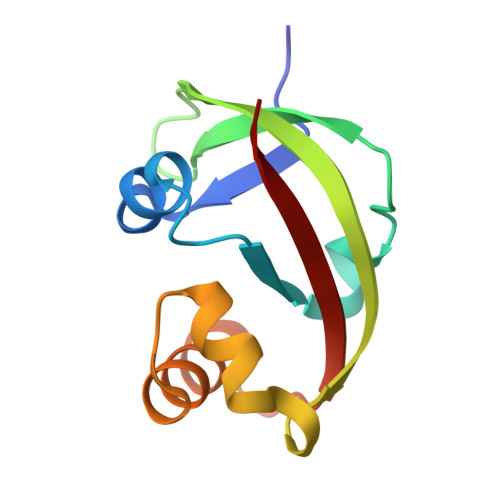
    9KYF, 9KYG, 9KYH, 9KYI, 9KYJ, 9KYK, 9KYL

  • PubMed Abstract: 

    ASC-1 homology (ASCH) domain family proteins are believed to play essential roles in RNA metabolism, but detailed structural and functional information is limited. Research has shown that the E. coli enzyme YqfB, which contains an ASCH domain, has amidohydrolase activity, converting N 4 -acetylcytidine (ac 4 C) RNA nucleoside into cytidine. Here, we present the crystal structures of EcYqfB both in its unbound state and bound to a substrate. Our analysis reveals how the substrate interacts with the enzyme, offering insights into its catalytic mechanism. In vivo experiments further show that deleting EcYqfB does not change overall ac 4 C levels across various RNA types, indicating that EcYqfB specifically functions in ac 4 C nucleoside metabolism. We also determined the structures of two homologous proteins: mouse EOLA1 and the human TRIP4-ASCH domain, highlighting differences in their substrate preferences. These findings offer important insights for future research into the structure and function of the ASCH domain protein family.


  • Organizational Affiliation
    • China-New Zealand Joint Laboratory on Biomedicine and Health, Institute of Drug Discovery, Guangzhou Institutes of Biomedicine and Health (GIBH), Chinese Academy of Sciences (CAS), Guangzhou 510530, China.

Macromolecule Content 

  • Total Structure Weight: 23.84 kDa 
  • Atom Count: 1,988 
  • Modeled Residue Count: 203 
  • Deposited Residue Count: 206 
  • Unique protein chains: 1

Macromolecules

Find similar proteins by:|  3D Structure
Entity ID: 1
MoleculeChains  Sequence LengthOrganismDetailsImage
N(4)-acetylcytidine amidohydrolase
A, B
103Escherichia coli BL21(DE3)Mutation(s): 0 
Gene Names: ECBD_0837
EC: 3.5.1.135
UniProt
Find proteins for P67603 (Escherichia coli (strain K12))
Explore P67603 
Go to UniProtKB:  P67603
Entity Groups
Sequence Clusters30% Identity50% Identity70% Identity90% Identity95% Identity100% Identity
UniProt GroupP67603
Sequence Annotations
Expand
Reference Sequence

Small Molecules

Ligands 1 Unique
IDChains Name / Formula / InChI Key2D Diagram3D Interactions
CA

Query on CA


Download:Ideal Coordinates CCD File
C [auth A]CALCIUM ION
Ca
BHPQYMZQTOCNFJ-UHFFFAOYSA-N

Experimental Data & Validation

Experimental Data

  • Method: X-RAY DIFFRACTION
  • Resolution: 1.55 Å
  • R-Value Free:  0.245 (Depositor), 0.244 (DCC) 
  • R-Value Work:  0.201 (Depositor), 0.202 (DCC) 
  • R-Value Observed: 0.203 (Depositor) 
Space Group: C 1 2 1
Unit Cell:
Length ( Å )Angle ( ˚ )
a = 108.306α = 90
b = 30.55β = 93.897
c = 61.949γ = 90
Software Package:
Software NamePurpose
PHENIXrefinement
autoPROCdata reduction
SCALAdata scaling
PHASERphasing

Structure Validation

View Full Validation Report


Entry History 

& Funding Information

Deposition Data


Funding OrganizationLocationGrant Number
Not funded--

Revision History  (Full details and data files)

  • Version 1.0: 2025-08-20
    Type: Initial release
  • Version 1.1: 2025-09-24
    Changes: Database references
  • Version 1.2: 2025-12-24
    Changes: Database references