


Crystal structure of proteinase K from Engyodontium album
Sugahara, M., Maki-Yonekura, S., Yonekura, K.To be published.
Experimental Data Snapshot
wwPDB Validation 3D Report Full Report

Entity ID: 1 | |||||
|---|---|---|---|---|---|
| Molecule | Chains | Sequence Length | Organism | Details | Image |
| Proteinase K | 279 | Parengyodontium album | Mutation(s): 0 Gene Names: PROK EC: 3.4.21.64 | 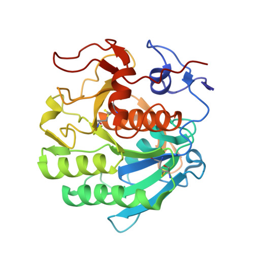 | |
UniProt | |||||
Entity Groups | |||||
| Sequence Clusters | 30% Identity50% Identity70% Identity90% Identity95% Identity100% Identity | ||||
| UniProt Group | P06873 | ||||
Sequence AnnotationsExpand | |||||
Reference Sequence | |||||
| Ligands 2 Unique | |||||
|---|---|---|---|---|---|
| ID | Chains | Name / Formula / InChI Key | 2D Diagram | 3D Interactions | |
| EU (Subject of Investigation/LOI) Download:Ideal Coordinates CCD File | B [auth A], C [auth A] | EUROPIUM ION Eu MGVUQZZTJGLWJV-UHFFFAOYSA-N |  | ||
| NO3 (Subject of Investigation/LOI) Download:Ideal Coordinates CCD File | D [auth A] | NITRATE ION N O3 NHNBFGGVMKEFGY-UHFFFAOYSA-N |  | ||
| Length ( Å ) | Angle ( ˚ ) |
|---|---|
| a = 68.7 | α = 90 |
| b = 68.7 | β = 90 |
| c = 108.98 | γ = 90 |
| Software Name | Purpose |
|---|---|
| PHENIX | refinement |
| CrystFEL | data scaling |
| SHELXCD | phasing |
| Funding Organization | Location | Grant Number |
|---|---|---|
| Not funded | -- |


