9KLU | pdb_00009klu

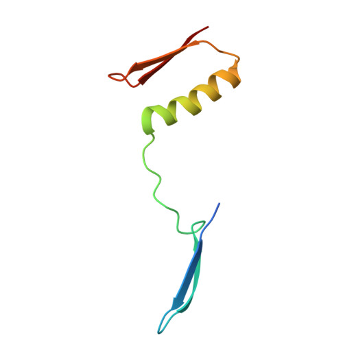
Crystal structure of a Streptococcal protein G B1 mutant


Experimental Data Snapshot

  • Method: X-RAY DIFFRACTION
  • Resolution: 2.91 Å
  • R-Value Free: 
    0.266 (Depositor), 0.264 (DCC) 
  • R-Value Work: 
    0.234 (Depositor), 0.235 (DCC) 
  • R-Value Observed: 
    0.236 (Depositor) 

Starting Model: experimental
View more details

wwPDB Validation 3D Report Full Report

Validation slider image for 9KLU

This is version 1.0 of the entry. See complete history

Literature

Regulating the orientation of homobivalent small binders through 3D domain-swapping design.

Shiga, S.Watanabe, H.Makabe, K.Honda, S.

(2025) J Biological Chem 302: 111053-111053

  • DOI: https://doi.org/10.1016/j.jbc.2025.111053
  • Primary Citation Related Structures: 
    9KL7, 9KLS, 9KLT, 9KLU

  • PubMed Abstract: 

    Homobivalent small binders represent bioactive dimeric proteins constructed by linking nanobodies or other small binders. As binding domain orientation in these dimeric binders affects binding affinity, expanding the orientation regulation techniques is crucial for optimizing their performance. Here, we report a 3D domain-swapping design as a genetic engineering approach to regulate the orientation of homobivalent small binders. We aimed to regulate B1 domain orientation of protein G as a model binder. By inserting single cysteine-containing polyproline sequences into loops located at the C- and N-terminal sides of the tertiary structure, we successfully designed 3D domain-swapped dimers with tail-to-tail and head-to-head orientations. Notably, the head-to-head-oriented dimer demonstrated potential as a functional homobivalent small binder. Our results highlight the applicability of the 3D domain-swapping design for regulating the orientation of homobivalent small binders and establish a foundation for developing 3D domain-swapped dimers.


  • Organizational Affiliation
    • Molecular Biosystems Research Institute, National Institute of Advanced Industrial Science and Technology (AIST), Tsukuba, Ibaraki, Japan; Graduate School of Science and Engineering, Yamagata University, Yonezawa, Yamagata, Japan.

Macromolecule Content 

  • Total Structure Weight: 14.73 kDa 
  • Atom Count: 980 
  • Modeled Residue Count: 126 
  • Deposited Residue Count: 134 
  • Unique protein chains: 1

Macromolecules

Find similar proteins by:|  3D Structure
Entity ID: 1
MoleculeChains  Sequence LengthOrganismDetailsImage
Protein LG
A, B
67Finegoldia magnaMutation(s): 4 
UniProt
Find proteins for Q53291 (Finegoldia magna)
Explore Q53291 
Go to UniProtKB:  Q53291
Entity Groups
Sequence Clusters30% Identity50% Identity70% Identity90% Identity95% Identity100% Identity
UniProt GroupQ53291
Sequence Annotations
Expand
Reference Sequence

Experimental Data & Validation

Experimental Data

  • Method: X-RAY DIFFRACTION
  • Resolution: 2.91 Å
  • R-Value Free:  0.266 (Depositor), 0.264 (DCC) 
  • R-Value Work:  0.234 (Depositor), 0.235 (DCC) 
  • R-Value Observed: 0.236 (Depositor) 
Space Group: P 21 21 21
Unit Cell:
Length ( Å )Angle ( ˚ )
a = 43.506α = 90
b = 75.058β = 90
c = 96.925γ = 90
Software Package:
Software NamePurpose
PHENIXrefinement
XDSdata reduction
XDSdata scaling
PHASERphasing
Cootmodel building

Structure Validation

View Full Validation Report


Entry History 

& Funding Information

Deposition Data


Funding OrganizationLocationGrant Number
Other governmentJapan--

Revision History  (Full details and data files)

  • Version 1.0: 2026-01-28
    Type: Initial release