


Structure of PfENT1(S143C,S382C) with inosine
Chen, X.Y., Li, J.L., Wang, X., Deng, D.To be published.
Experimental Data Snapshot
wwPDB Validation 3D Report Full Report

Entity ID: 1 | |||||
|---|---|---|---|---|---|
| Molecule | Chains | Sequence Length | Organism | Details | Image |
| Nucleoside transporter 1 | 384 | Plasmodium falciparum 3D7 | Mutation(s): 2 Gene Names: NT1, PF3D7_1347200 | 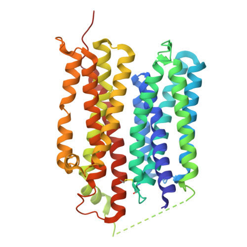 | |
UniProt | |||||
Entity Groups | |||||
| Sequence Clusters | 30% Identity50% Identity70% Identity90% Identity95% Identity100% Identity | ||||
| UniProt Group | Q8IDM6 | ||||
Sequence AnnotationsExpand | |||||
Reference Sequence | |||||
| Ligands 1 Unique | |||||
|---|---|---|---|---|---|
| ID | Chains | Name / Formula / InChI Key | 2D Diagram | 3D Interactions | |
| NOS (Subject of Investigation/LOI) Download:Ideal Coordinates CCD File | B [auth A] | INOSINE C10 H12 N4 O5 UGQMRVRMYYASKQ-KQYNXXCUSA-N |  | ||
| Task | Software Package | Version |
|---|---|---|
| MODEL REFINEMENT | PHENIX | |
| Funding Organization | Location | Grant Number |
|---|---|---|
| National Natural Science Foundation of China (NSFC) | China | 3217110258 |


