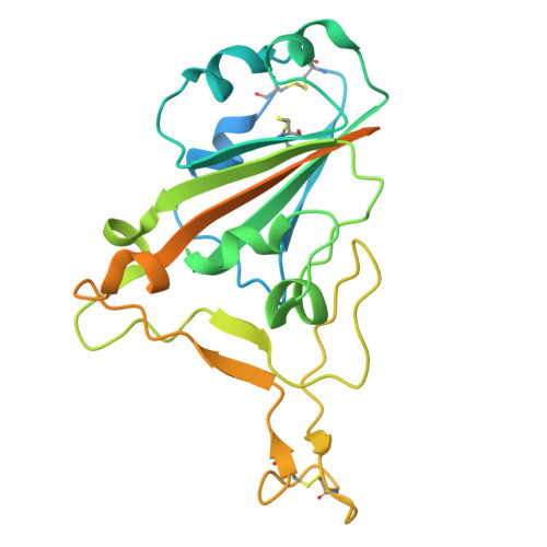
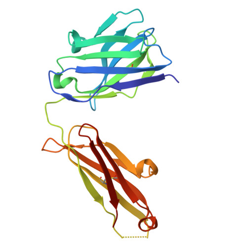
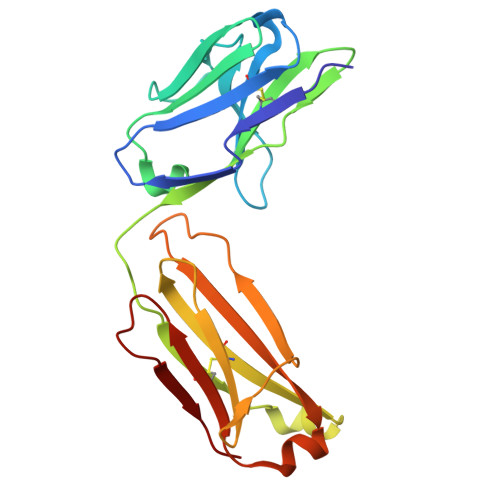
Mechanistic studies of viral neutralization typically prioritize high-affinity antibodies, relegating low-affinity binders to the sidelines. P5‑1C8, a Class 1 SARS-CoV-2 antibody that exemplifies this underexplored "low‑affinity yet high‑potency" phenotype is reported, retaining strong neutralization of Omicron JN.1 despite markedly weakened trimer binding (K D = 225 nM; IC 50 = 0.06 nM). Structural and biophysical analyses reveal that P5-1C8 engages WT and BA.1 spikes through canonical intra-spike bivalency, but with JN.1 it induces aggregation. Using virion-like nanoparticles displaying multiple spikes, it is shown that IgG remains bound with no detectable dissociation and triggers pronounced aggregation. Coarse-grained molecular dynamics delineate the stepwise pathway in which weak IgG-spike contacts seed aggregation via transient inter-spike bridging. Together, these findings establish the first mechanistic framework demonstrating how weak-binding antibodies can nonetheless achieve potent neutralization through higher-order aggregation, thereby expanding the conceptual landscape of antibody function and opening new directions for antibody evaluation and design.
Organizational Affiliation:
National Cancer Center/National Clinical Research Center for Cancer/Cancer Hospital, Chinese Academy of Medical Sciences and Peking Union Medical College, Beijing, 100021, China.
Laboratory of Nanosystem and Hierarchical Fabrication, National Center for Nanoscience and Technology, Beijing, 100190, China.
Research Unit of Nanoscience and Technology, Chinese Academy of Medical Sciences, Beijing, 100730, China.
Comprehensive AIDS Research Center, Pandemic Research Alliance Unit, Center for Infection Biology, School of Basic Medical Sciences, Tsinghua University, Beijing, 100084, China.
State Key Laboratory of Chemical Engineering and Low-carbon Technology, Department of Chemical Engineering and Low-carbon Technology, Tsinghua University, Beijing, 100084, China.
CAS Key Laboratory for Biological Effects of Nanomaterials and Nanosafety, National Center for Nanoscience and Technology, Chinese Academy of Sciences, Beijing, 100190, China.
College of Ecology, Lanzhou University, Lanzhou, 730000, China.
University of Chinese Academy of Sciences, Beijing, 100049, China.
The Salk Institute of Biological Sciences, La Jolla, CA, 92037, USA.
The Ministry of Education Key Laboratory of Protein Science, Beijing Frontier Research Center for Biological Structure, School of Life Sciences, Tsinghua University, Beijing, 100084, China.
CAS Key Laboratory of Colloid, Interface and Chemical Thermodynamics, Institute of Chemistry, Chinese Academy of Sciences, Beijing, 100190, China.
MOE Engineering Research Center of Advanced Rare Earth Materials, Department of Chemistry, Tsinghua University, Beijing, 100084, China.
Shanghai Institute of Infectious Disease and Biosecurity, Shanghai Medical College, Fudan University, Shanghai, 200032, China.
Centre for Virology, Vaccinology and Therapeutics, Department of Microbiology, Li Ka Shing Faculty of Medicine, The University of Hong Kong, Hong Kong SAR, 999077, China.
The technology center for protein science, Tsinghua University, Beijing, 100084, China.
Department of Applied Biology and Chemical Technology, The Hong Kong Polytechnic University, Hong Hum, Kowloon, 999077, Hong Kong.
PolyU Shenzhen Research Institute, Shenzhen, 518057, China.
Zhejiang Provincial Key Laboratory for Cancer Molecular Cell Biology, Life Sciences Institute, Zhejiang University, Hangzhou, 310058, China.
Institute of Bio-Architecture and Bio-Interactions, Shenzhen Medical Academy of Research and Translation, Shenzhen, Guangdong, 518107, China.
CAS Key Laboratory for Biomedical Effects of Nanomaterials and Nanosafety and CAS Center for Excellence in Nanoscience, Beijing, 100190, China.
Department of Gastrointestinal Oncology Key Laboratory of Carcinogenesis and Translational Research (Ministry of Education), Beijing, 100142, China.
Institute of Biopharmaceutical and Health Engineering, Tsinghua Shenzhen International Graduate School, Tsinghua University, Shenzhen, 518055, China.
Institute of Biomedical Health Technology and Engineering, Shenzhen Bay Laboratory, Shenzhen, 518132, China.








 Download Mendeley