Electrostatic tuning of the pyridoxal-5'-phosphate cofactor site defines pH dependence in type I cystathionine beta-lyases.
Liu, Y., Li, X., Li, J., Liu, J., Peng, Y., Gao, Y., Zhang, Y., Jia, Z., Kang, K., Yin, A., Ma, C., Yang, Y., Yang, C.(2026) J Biological Chem 302: 111469-111469
- PubMed: 42001944 Search on PubMedSearch on PubMed Central
- DOI: https://doi.org/10.1016/j.jbc.2026.111469
- Primary Citation Related Structures:
8Y54, 9JVA, 9K71 - PubMed Abstract:
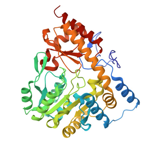
Thiosulfinates, commonly known as "plant antibiotics", exhibit broad-spectrum antimicrobial activity, but are inherently unstable under alkaline conditions. Thus, the development of biocatalysts that maintain high efficiency at acidic pH is crucial. In this study, we determined the structures of two patB gene-encoded cystathionine β-lyases from the fold-type I pyridoxal-5'-phosphate (PLP)-dependent family, BcPatB and LdPatB, which exhibit distinct pH dependencies in catalyzing the conversion of l-cysteine-S-conjugate sulfoxides to thiosulfinates. Structural comparison of these two PatBs revealed a residue pair located near the negatively charged phosphate group of the PLP cofactor, exhibiting distinct charge properties. By mutating this residue pair to enhance negative-negative charge interactions with the PLP phosphate, we generated the BcPatB mutant H263E/Q238M and LdPatB mutant H241M, both of which exhibited significantly improved activity at mildly acidic pH of 6.0. This strategy was subsequently applied to other fold-type I PLP-dependent enzymes, including MePatB, metC gene-encoded cystathionine β-lyase from Klebsiella pneumoniae (KpMetC), and the alanine aminotransferase from Escherichia coli K-12 (Eck-12AlaA), resulting in mutants with modified pH preferences. Constant-pH molecular dynamics simulations demonstrated that the modified electrostatic interactions between PLP and the residue pair play a key role in driving pH-dependent catalysis in these mutants. These findings suggest that this residue pair may function as a pH switch in certain fold-type I PLP-dependent enzymes, and that rational engineering of this position offers a promising strategy for tailoring enzymes to specific industrial pH conditions.
- State Key Laboratory of Microbial Technology, Institute of Microbial Technology, Shandong University, Qingdao, P. R. China.
Organizational Affiliation: