9JU4 | pdb_00009ju4

Crystal structure of the Thermotoga maritima cold shock protein mutant Est#13 in complex with AacEst


Experimental Data Snapshot

  • Method: X-RAY DIFFRACTION
  • Resolution: 2.00 Å
  • R-Value Free: 
    0.207 (Depositor), 0.210 (DCC) 
  • R-Value Work: 
    0.170 (Depositor), 0.173 (DCC) 
  • R-Value Observed: 
    0.171 (Depositor) 

Starting Model: experimental
View more details

wwPDB Validation   3D Report Full Report


Ligand Structure Quality Assessment 

Created with Raphaël 2.3.0Worse 01 BetterLigand structure goodness of fit to experimental dataBest fitted PMSClick on this verticalbar to view detailsBest fitted PEGClick on this verticalbar to view detailsBest fitted GOLClick on this verticalbar to view details

This is version 1.0 of the entry. See complete history


Literature

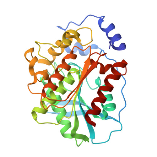
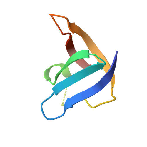
Heat-sterilizable antibody mimics designed on the cold shock protein scaffold from hyperthermophile Thermotoga maritima.

Amesaka, H.Tachibana, M.Hara, M.Toya, S.Nakagawa, H.Matsumura, H.Hirata, A.Fujihashi, M.Takano, K.Tanaka, S.I.

(2025) Protein Sci 34: e70018-e70018

  • DOI: https://doi.org/10.1002/pro.70018
  • Primary Citation of Related Structures:  
    9JU4

  • PubMed Abstract: 

    Antibodies and antibody mimics are extensively used in the pharmaceutical industry, where stringent safety standards are required. Implementing heat sterilization during or after the manufacturing process could help prevent contamination by viruses and bacteria. However, conventional antibodies and antibody mimics are not suitable for heat sterilization because they irreversibly denature at high temperatures. In this study, we focused on the refolding property of the cold shock protein from the hyperthermophile Thermotoga maritima (TmCSP), which denatures at elevated temperatures but regains its native structure upon re-cooling. We designed and constructed a mutant library of TmCSP in which amino acid residues in its three surface loops were diversified. From the library, mutant TmCSPs that bind to each of eight target proteins were selected by phage and yeast surface display methods. We confirmed that the secondary structure and binding affinity of all the selected mutants were restored after heat treatment followed by cooling. Additionally, freeze-drying did not impair their binding affinity. The crystal structure of a mutant TmCSP in complex with its target, the esterase from Alicyclobacillus acidocaldarius, revealed specific interactions between them. These results clearly demonstrate the feasibility of creating heat-sterilizable antibody mimics using TmCSP as a scaffold.


  • Organizational Affiliation
    • Graduate School of Life and Environmental Sciences, Kyoto Prefectural University, Kyoto, Japan.

Macromolecules
Find similar proteins by:  (by identity cutoff)  |  3D Structure
Entity ID: 1
MoleculeChains Sequence LengthOrganismDetailsImage
Alpha/beta hydrolase fold-3 domain protein310Alicyclobacillus acidocaldariusMutation(s): 0 
Gene Names: Aaci_2875
UniProt
Find proteins for C8WUR3 (Alicyclobacillus acidocaldarius subsp. acidocaldarius (strain ATCC 27009 / DSM 446 / BCRC 14685 / JCM 5260 / KCTC 1825 / NBRC 15652 / NCIMB 11725 / NRRL B-14509 / 104-IA))
Explore C8WUR3 
Go to UniProtKB:  C8WUR3
Entity Groups  
Sequence Clusters30% Identity50% Identity70% Identity90% Identity95% Identity100% Identity
UniProt GroupC8WUR3
Sequence Annotations
Expand
  • Reference Sequence
Find similar proteins by:  (by identity cutoff)  |  3D Structure
Entity ID: 2
MoleculeChains Sequence LengthOrganismDetailsImage
The Thermotoga maritima cold shock protein mutant binding to AacEst66Thermotoga maritimaMutation(s): 0 
UniProt
Find proteins for O54310 (Thermotoga maritima (strain ATCC 43589 / DSM 3109 / JCM 10099 / NBRC 100826 / MSB8))
Explore O54310 
Go to UniProtKB:  O54310
Entity Groups  
Sequence Clusters30% Identity50% Identity70% Identity90% Identity95% Identity100% Identity
UniProt GroupO54310
Sequence Annotations
Expand
  • Reference Sequence
Experimental Data & Validation

Experimental Data

  • Method: X-RAY DIFFRACTION
  • Resolution: 2.00 Å
  • R-Value Free:  0.207 (Depositor), 0.210 (DCC) 
  • R-Value Work:  0.170 (Depositor), 0.173 (DCC) 
  • R-Value Observed: 0.171 (Depositor) 
Space Group: P 1 2 1
Unit Cell:
Length ( Å )Angle ( ˚ )
a = 45.82α = 90
b = 46.65β = 93.99
c = 101.26γ = 90
Software Package:
Software NamePurpose
PHENIXrefinement
XDSdata reduction
XDSdata scaling
PHENIXphasing

Structure Validation

View Full Validation Report


Ligand Structure Quality Assessment 

Created with Raphaël 2.3.0Worse 01 BetterLigand structure goodness of fit to experimental dataBest fitted PMSClick on this verticalbar to view detailsBest fitted PEGClick on this verticalbar to view detailsBest fitted GOLClick on this verticalbar to view details

Entry History & Funding Information

Deposition Data


Funding OrganizationLocationGrant Number
Japan Society for the Promotion of Science (JSPS)Japan21K05386
Japan Society for the Promotion of Science (JSPS)Japan23H04559
Japan Society for the Promotion of Science (JSPS)Japan24K08717
Japan Society for the Promotion of Science (JSPS)Japan23KJ1820

Revision History  (Full details and data files)

  • Version 1.0: 2025-01-15
    Type: Initial release