9JT0 | pdb_00009jt0

Chito oligosaccharide deacetylase from vibrio campbellii (VhCOD) complex with Chitobiose


Experimental Data Snapshot

  • Method: X-RAY DIFFRACTION
  • Resolution: 2.40 Å
  • R-Value Free: 
    0.217 (Depositor), 0.218 (DCC) 
  • R-Value Work: 
    0.180 (Depositor), 0.182 (DCC) 
  • R-Value Observed: 
    0.181 (Depositor) 

Starting Model: experimental
View more details

wwPDB Validation 3D Report Full Report

Validation slider image for 9JT0

This is version 1.1 of the entry. See complete history

Literature

Structure and loop dynamics of a chitooligosaccharide deacetylase from the marine bacterium Vibrio campbellii (harveyi).

Pongnan, S.Robinson, R.C.Lampela, O.Juffer, A.Fukamizo, T.Suginta, W.

(2025) J Biological Chem 301: 110608-110608

  • DOI: https://doi.org/10.1016/j.jbc.2025.110608
  • Primary Citation Related Structures: 
    8YFP, 8YH4, 9JEN, 9JEO, 9JT0, 9JT8

  • PubMed Abstract: 

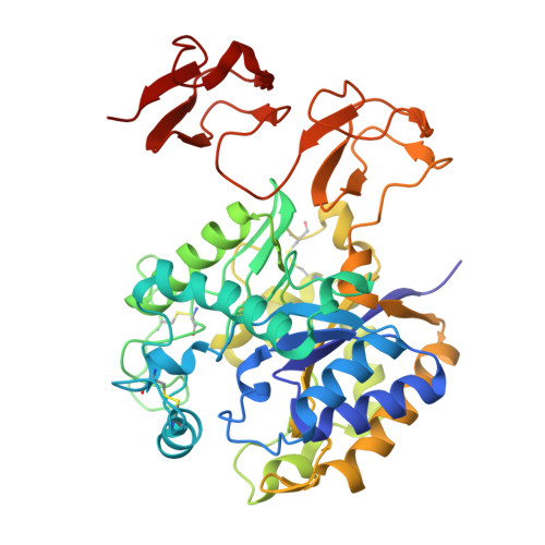
    A chitooligosaccharide deacetylase from Vibrio campbellii (formerly Vibrio harveyi) (VhCOD) belonging to the carbohydrate esterase family 4 catalyzes Zn 2+ -dependent deacetylation of a specific GlcNAc residue in chitooligosaccharides. It deacetylates chitobiose, (GlcNAc) 2 , to produce GlcNAc-GlcN following Michaelis-Menten kinetics. We elucidated the six crystal structures of wildtype VhCOD in ligand-free and -bound states with (GlcNAc) 2 (substrate), GlcNAc-GlcN (product), (GlcN) 2 (product analog), GlcNAc-GlcN-GlcNAc (product), or GlcNAc-GlcN-(GlcNAc) 2 (product). The structures of VhCOD comprise the carbohydrate esterase family 4 catalytic domain and the two CBM12 carbohydrate-binding domains, similar to the COD homologs from Vibrio cholerae and Vibrio parahaemolyticus. The catalytic site, where a Zn 2+ ion is coordinated with the His-His-Asp triad and three water molecules, is surrounded by six loops (L1-L6). Comparison between the ligand-free and various bound structures uncovered full catalytic cycle, including the product release in company with a large conformational change in L4. Molecular dynamics simulation based on the crystal structures provided further insights into the loop fluctuations, which are proposed to be involved in the catalytic reaction.


  • Organizational Affiliation
    • School of Biomolecular Science and Engineering (BSE), Vidyasirimedhi Institute of Science and Technology (VISTEC), Rayong, Thailand.

Macromolecule Content 

  • Total Structure Weight: 134.73 kDa 
  • Atom Count: 9,861 
  • Modeled Residue Count: 1,211 
  • Deposited Residue Count: 1,218 
  • Unique protein chains: 1

Macromolecules

Find similar proteins by:|  3D Structure
Entity ID: 1
MoleculeChains  Sequence LengthOrganismDetailsImage
NodB homology domain-containing protein
A, B, C
406Vibrio campbellii ATCC BAA-1116Mutation(s): 0 
Gene Names: VIBHAR_03626
UniProt
Find proteins for A7MSF4 (Vibrio campbellii (strain ATCC BAA-1116))
Explore A7MSF4 
Go to UniProtKB:  A7MSF4
Entity Groups
Sequence Clusters30% Identity50% Identity70% Identity90% Identity95% Identity100% Identity
UniProt GroupA7MSF4
Sequence Annotations
Expand
Reference Sequence

Oligosaccharides

Help  
Entity ID: 2
MoleculeChains Length2D Diagram GlycosylationD Interactions
2-acetamido-2-deoxy-beta-D-glucopyranose-(1-4)-2-acetamido-2-deoxy-beta-D-glucopyranoseD [auth E]2N/A
Glycosylation Resources
GlyTouCan: G42666HT
GlyCosmos: G42666HT
GlyGen: G42666HT

Experimental Data & Validation

Experimental Data

  • Method: X-RAY DIFFRACTION
  • Resolution: 2.40 Å
  • R-Value Free:  0.217 (Depositor), 0.218 (DCC) 
  • R-Value Work:  0.180 (Depositor), 0.182 (DCC) 
  • R-Value Observed: 0.181 (Depositor) 
Space Group: P 31 1 2
Unit Cell:
Length ( Å )Angle ( ˚ )
a = 107.852α = 90
b = 107.852β = 90
c = 260.618γ = 120
Software Package:
Software NamePurpose
PHENIXrefinement
HKL-2000data scaling
XDSdata reduction
PHENIXphasing

Structure Validation

View Full Validation Report


Entry History 

& Funding Information

Deposition Data


Funding OrganizationLocationGrant Number
Vidyasirimedhi Institute of Science and Technology (VISTEC)Thailand--

Revision History  (Full details and data files)

  • Version 1.0: 2025-10-01
    Type: Initial release
  • Version 1.1: 2025-10-22
    Changes: Database references