9J93 | pdb_00009j93

Native GluA1/GluA4 ATD dimer binding with 1D8 and 11B8


Experimental Data Snapshot

  • Method: ELECTRON MICROSCOPY
  • Resolution: 3.54 Å
  • Aggregation State: PARTICLE 
  • Reconstruction Method: SINGLE PARTICLE 

wwPDB Validation 3D Report Full Report

Validation slider image for 9J93

This is version 1.2 of the entry. See complete history

Literature

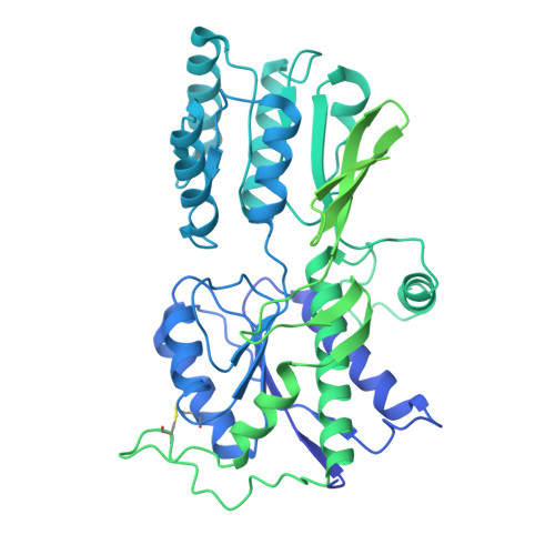
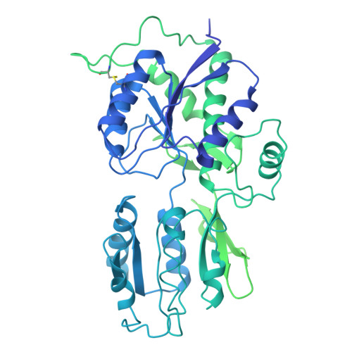
Assembly and gating mechanism of native AMPA receptors from the cerebellum.

Li, X.Li, R.Wei, Y.Chen, J.Zhao, J.Zhao, J.Wang, W.Li, N.Wang, L.Hu, T.Dong, Y.Zhu, Y.Wei, C.Li, L.Zhang, W.Huang, Z.Zhao, Y.

(2026) Cell Res 

  • DOI: https://doi.org/10.1038/s41422-026-01234-8
  • Primary Citation Related Structures: 
    9J92, 9J93, 9J94, 9J95, 9X57, 9X58, 9XJL, 9XJM

  • PubMed Abstract: 

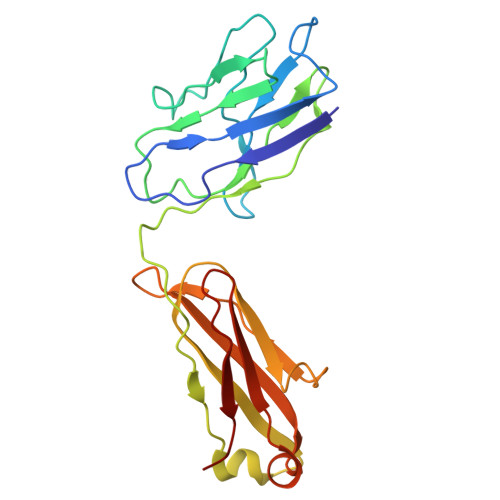
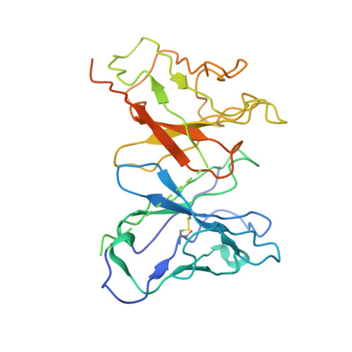
    AMPA receptors (AMPARs) mediate the majority of fast excitatory synaptic transmission throughout the central nervous system. Calcium-permeable AMPARs and GluA4-containing receptors are critical for cerebellar functions, such as motor learning, associative memory, auditory processing, and synaptic plasticity. In contrast to the well-characterized, predominantly GluA2-containing AMPARs of the hippocampus and cortex, cerebellar AMPARs contain a higher proportion of GluA4 and remain poorly understood. Here, we generated a highly GluA4-specific antibody. Using this antibody in combination with antibodies specifically recognizing GluA1 and GluA2, we purified native AMPARs and determined the subunit compositions of both calcium-impermeable and calcium-permeable native AMPARs in the cerebellum. The isolated cerebellar AMPARs that contained both GluA1 and GluA4 were calcium-permeable, with GluA4 occupying mainly the B/D positions, GluA1 occupying the A/C positions, and the complex associated primarily with cornichon 3 (CNIH3). We determined the structures of the complex in distinct functional states, including the resting, active, and desensitized states, and characterized the conformational transitions that underlie its activity. During desensitization, the receptor adopts a pseudo-4-fold configuration of the ligand-binding domain layer, which may be important for its functional properties. This study provides a blueprint for the subunit compositions of AMPARs in the cerebellum and clarifies the gating mechanism of the calcium-permeable native AMPAR A1A4 -CNIH3 complex, providing significant insight into AMPAR-mediated synaptic transmission in the cerebellum.


  • Organizational Affiliation
    • Key Laboratory of Biomacromolecules (CAS), National Laboratory of Biomacromolecules, CAS Center for Excellence in Biomacromolecules, Institute of Biophysics, Chinese Academy of Sciences, Beijing, China.

Macromolecule Content 

  • Total Structure Weight: 269.68 kDa 
  • Atom Count: 10,138 
  • Modeled Residue Count: 1,412 
  • Deposited Residue Count: 2,501 
  • Unique protein chains: 4

Macromolecules

Find similar proteins by:|  3D Structure
Entity ID: 1
MoleculeChains  Sequence LengthOrganismDetailsImage
Glutamate receptorA [auth B]884Sus scrofaMutation(s): 0 
UniProt
Find proteins for I3L8N9 (Sus scrofa)
Explore I3L8N9 
Go to UniProtKB:  I3L8N9
Entity Groups
Sequence Clusters30% Identity50% Identity70% Identity90% Identity95% Identity100% Identity
UniProt GroupI3L8N9
Glycosylation
Glycosylation Sites: 1
Sequence Annotations
Expand
Reference Sequence
Find similar proteins by:|  3D Structure
Entity ID: 2
MoleculeChains  Sequence LengthOrganismDetailsImage
Glutamate receptorB [auth A]906Sus scrofaMutation(s): 0 
UniProt
Find proteins for A0A286ZS63 (Sus scrofa)
Explore A0A286ZS63 
Go to UniProtKB:  A0A286ZS63
Entity Groups
Sequence Clusters30% Identity50% Identity70% Identity90% Identity95% Identity100% Identity
UniProt GroupA0A286ZS63
Glycosylation
Glycosylation Sites: 3
Sequence Annotations
Expand
Reference Sequence
Find similar proteins by:|  3D Structure
Entity ID: 3
MoleculeChains  Sequence LengthOrganismDetailsImage
1D8 FabC [auth H],
D [auth L]
218Sus scrofaMutation(s): 0 
Find similar proteins by:|  3D Structure
Entity ID: 4
MoleculeChains  Sequence LengthOrganismDetailsImage
11B8 scFVE [auth C]275Mus musculusMutation(s): 0 

Oligosaccharides

Help  
Entity ID: 5
MoleculeChains Length2D Diagram GlycosylationD Interactions
2-acetamido-2-deoxy-beta-D-glucopyranose-(1-4)-2-acetamido-2-deoxy-beta-D-glucopyranoseF [auth D],
G [auth E],
H [auth F],
I [auth G],
J [auth I]
2N-Glycosylation
Glycosylation Resources
GlyTouCan: G42666HT
GlyCosmos: G42666HT
GlyGen: G42666HT

Small Molecules

Ligands 1 Unique
IDChains Name / Formula / InChI Key2D Diagram3D Interactions
NAG

Query on NAG


Download:Ideal Coordinates CCD File
K [auth A]2-acetamido-2-deoxy-beta-D-glucopyranose
C8 H15 N O6
OVRNDRQMDRJTHS-FMDGEEDCSA-N

Experimental Data & Validation

Experimental Data

  • Method: ELECTRON MICROSCOPY
  • Resolution: 3.54 Å
  • Aggregation State: PARTICLE 
  • Reconstruction Method: SINGLE PARTICLE 
EM Software:
TaskSoftware PackageVersion
MODEL REFINEMENTPHENIX

Structure Validation

View Full Validation Report


Entry History 

& Funding Information

Deposition Data


Funding OrganizationLocationGrant Number
National Natural Science Foundation of China (NSFC)China92157102

Revision History  (Full details and data files)

  • Version 1.0: 2026-03-04
    Type: Initial release
  • Version 1.1: 2026-03-25
    Changes: Data collection, Database references
  • Version 1.2: 2026-04-01
    Changes: Data collection, Database references