9J4Y | pdb_00009j4y

Crystal Structure of the L322F mutant of Omega Transaminase TA_2799 from Pseudomonas putida KT2440


Experimental Data Snapshot

  • Method: X-RAY DIFFRACTION
  • Resolution: 2.50 Å
  • R-Value Free: 
    0.233 (Depositor), 0.234 (DCC) 
  • R-Value Work: 
    0.194 (Depositor), 0.200 (DCC) 
  • R-Value Observed: 
    0.196 (Depositor) 

Starting Model: experimental
View more details

wwPDB Validation 3D Report Full Report

Validation slider image for 9J4Y

This is version 1.2 of the entry. See complete history

Literature

Structural insights and rational design of Pseudomonasputida KT2440 omega transaminases for enhanced biotransformation of (R)-PAC to (1R, 2S)-Norephedrine.

Das, P.Noronha, S.Bhaumik, P.

(2025) J Biological Chem 301: 110289-110289

  • DOI: https://doi.org/10.1016/j.jbc.2025.110289
  • Primary Citation Related Structures: 
    9J2K, 9J4Y, 9J4Z, 9J50

  • PubMed Abstract: 

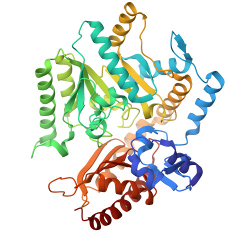
    Omega transaminases (ω-TAs) can mediate the chiral amination of several unnatural substrates without the requirement of an α-COOH group and are highly relevant in the production of several pharmaceutical intermediates of commercial interest. Development of better variants of ω-TAs is hence essential for biotransformation of unnatural substrates. We studied the active site architecture of the wild-type ω-TAs, to engineer enzymes that enhance the biotransformation of (R)-phenylacetylcarbinol to (1R, 2S)-norephedrine. Two such ω-TAs (TA_5182 and TA_2799) from P. putida KT2440 strain were overexpressed and purified as recombinant proteins. Crystal structures of TA_5182 were solved in two conformations, and significant movements of two highly flexible loops were observed in these different states. The TA_2799 structure was determined as a complex with the cofactor pyridoxal 5'-phosphate (PLP) covalently bound to the catalytic K286 as an internal aldimine. Enzyme assays indicated that TA_2799 required a four-fold higher cofactor concentration than TA_5182 to achieve satisfactory biotransformation of (R)-PAC. A key mutation of L322F in TA_2799 drastically reduced (∼8-fold) the cofactor dependency of the TA_2799_L322F mutant enzyme, and the mutant remained active for 96 h at 30°C. The crystal structure of the mutant enzyme revealed a key asparagine residue that mediates a hydrogen bonding network at the dimeric interface of the enzyme and is absent in TA_5182. The TA_5182_G119N mutant also showed enhanced cofactor affinity. The results of our studies will help generate Pseudomonad ω-TAs and ω-TAs from other organisms with high efficiency for asymmetric synthesis, for further applications in large-scale biotransformation processes.


  • Organizational Affiliation
    • Department of Biosciences and Bioengineering, Indian Institute of Technology Bombay, Powai, Mumbai-400076, India.

Macromolecule Content 

  • Total Structure Weight: 401.32 kDa 
  • Atom Count: 28,374 
  • Modeled Residue Count: 3,637 
  • Deposited Residue Count: 3,672 
  • Unique protein chains: 1

Macromolecules

Find similar proteins by:|  3D Structure
Entity ID: 1
MoleculeChains  Sequence LengthOrganismDetailsImage
Aminotransferase, class III
A, B, C, D, E
A, B, C, D, E, F, G, H
459Pseudomonas putida KT2440Mutation(s): 1 
Gene Names: PP_2799
EC: 2.6.1.113
UniProt
Find proteins for Q9I6J2 (Pseudomonas aeruginosa (strain ATCC 15692 / DSM 22644 / CIP 104116 / JCM 14847 / LMG 12228 / 1C / PRS 101 / PAO1))
Explore Q9I6J2 
Go to UniProtKB:  Q9I6J2
Entity Groups
Sequence Clusters30% Identity50% Identity70% Identity90% Identity95% Identity100% Identity
UniProt GroupQ9I6J2
Sequence Annotations
Expand
Reference Sequence

Small Molecules

Ligands 4 Unique
IDChains Name / Formula / InChI Key2D Diagram3D Interactions
PEG

Query on PEG


Download:Ideal Coordinates CCD File
DA [auth G],
I [auth A],
R [auth C],
S [auth C]
DI(HYDROXYETHYL)ETHER
C4 H10 O3
MTHSVFCYNBDYFN-UHFFFAOYSA-N
SO4

Query on SO4


Download:Ideal Coordinates CCD File
AA [auth E]
CA [auth F]
EA [auth G]
FA [auth G]
HA [auth H]
AA [auth E],
CA [auth F],
EA [auth G],
FA [auth G],
HA [auth H],
L [auth A],
P [auth B],
Q [auth B],
U [auth C],
X [auth D],
Y [auth D]
SULFATE ION
O4 S
QAOWNCQODCNURD-UHFFFAOYSA-L
GOL

Query on GOL


Download:Ideal Coordinates CCD File
V [auth C]GLYCEROL
C3 H8 O3
PEDCQBHIVMGVHV-UHFFFAOYSA-N
EDO

Query on EDO


Download:Ideal Coordinates CCD File
BA [auth F]
GA [auth H]
J [auth A]
K [auth A]
M [auth B]
BA [auth F],
GA [auth H],
J [auth A],
K [auth A],
M [auth B],
N [auth B],
O [auth B],
T [auth C],
W [auth D],
Z [auth E]
1,2-ETHANEDIOL
C2 H6 O2
LYCAIKOWRPUZTN-UHFFFAOYSA-N

Experimental Data & Validation

Experimental Data

  • Method: X-RAY DIFFRACTION
  • Resolution: 2.50 Å
  • R-Value Free:  0.233 (Depositor), 0.234 (DCC) 
  • R-Value Work:  0.194 (Depositor), 0.200 (DCC) 
  • R-Value Observed: 0.196 (Depositor) 
Space Group: P 1 21 1
Unit Cell:
Length ( Å )Angle ( ˚ )
a = 119.16α = 90
b = 121.44β = 111.88
c = 147.41γ = 90
Software Package:
Software NamePurpose
REFMACrefinement
XDSdata reduction
XSCALEdata scaling
MoRDaphasing

Structure Validation

View Full Validation Report


Entry History 

& Funding Information

Deposition Data


Funding OrganizationLocationGrant Number
Not funded--

Revision History  (Full details and data files)

  • Version 1.0: 2025-06-04
    Type: Initial release
  • Version 1.1: 2025-06-11
    Changes: Database references
  • Version 1.2: 2025-07-09
    Changes: Database references