9IVV | pdb_00009ivv

Crystal structure of human secretory glutaminyl cyclase in complex with the inhibitor 3-((2-(1H-imidazol-5-yl)ethyl)carbamoyl)-4-amino-1,2,5-oxadiazole 2-oxide (compound 13)


Experimental Data Snapshot

  • Method: X-RAY DIFFRACTION
  • Resolution: 2.96 Å
  • R-Value Free: 
    0.255 (Depositor), 0.254 (DCC) 
  • R-Value Work: 
    0.206 (Depositor), 0.212 (DCC) 
  • R-Value Observed: 
    0.211 (Depositor) 

Starting Model: experimental
View more details

wwPDB Validation 3D Report Full Report

Validation slider image for 9IVV

Ligand Structure Quality Assessment 


This is version 1.1 of the entry. See complete history

Literature

Knowledge-guided diffusion model for 3D ligand-pharmacophore mapping.

Yu, J.L.Zhou, C.Ning, X.L.Mou, J.Meng, F.B.Wu, J.W.Chen, Y.T.Tang, B.D.Liu, X.G.Li, G.B.

(2025) Nat Commun 16: 2269-2269

  • DOI: https://doi.org/10.1038/s41467-025-57485-3
  • Primary Citation Related Structures: 
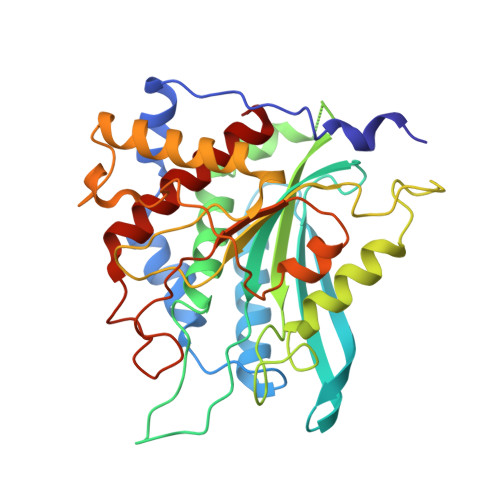
    9ISD, 9IVV

  • PubMed Abstract: 

    Pharmacophores are abstractions of essential chemical interaction patterns, holding an irreplaceable position in drug discovery. Despite the availability of many pharmacophore tools, the adoption of deep learning for pharmacophore-guided drug discovery remains relatively rare. We herein propose a knowledge-guided diffusion framework for 'on-the-fly' 3D ligand-pharmacophore mapping, named DiffPhore. It leverages ligand-pharmacophore matching knowledge to guide ligand conformation generation, meanwhile utilizing calibrated sampling to mitigate the exposure bias of the iterative conformation search process. By training on two self-established datasets of 3D ligand-pharmacophore pairs, DiffPhore achieves state-of-the-art performance in predicting ligand binding conformations, surpassing traditional pharmacophore tools and several advanced docking methods. It also manifests superior virtual screening power for lead discovery and target fishing. Using DiffPhore, we successfully identify structurally distinct inhibitors for human glutaminyl cyclases, and their binding modes are further validated through co-crystallographic analysis. We believe this work will advance the AI-enabled pharmacophore-guided drug discovery techniques.


  • Organizational Affiliation
    • Key Laboratory of Drug Targeting and Drug Delivery System of Ministry of Education, Department of Medicinal Chemistry, West China School of Pharmacy, Sichuan University, Chengdu, Sichuan, China.

Macromolecule Content 

  • Total Structure Weight: 38.46 kDa 
  • Atom Count: 2,701 
  • Modeled Residue Count: 323 
  • Deposited Residue Count: 329 
  • Unique protein chains: 1

Macromolecules

Find similar proteins by:|  3D Structure
Entity ID: 1
MoleculeChains  Sequence LengthOrganismDetailsImage
Glutaminyl-peptide cyclotransferase329Homo sapiensMutation(s): 0 
Gene Names: QPCT
EC: 2.3.2.5
UniProt & NIH Common Fund Data Resources
Find proteins for Q16769 (Homo sapiens)
Explore Q16769 
Go to UniProtKB:  Q16769
PHAROS:  Q16769
GTEx:  ENSG00000115828 
Entity Groups
Sequence Clusters30% Identity50% Identity70% Identity90% Identity95% Identity100% Identity
UniProt GroupQ16769
Sequence Annotations
Expand
Reference Sequence

Small Molecules

Ligands 5 Unique
IDChains Name / Formula / InChI Key2D Diagram3D Interactions
A1D94
(Subject of Investigation/LOI)

Query on A1D94


Download:Ideal Coordinates CCD File
C [auth A]4-azanyl-~{N}-[2-(1~{H}-imidazol-4-yl)ethyl]-2-oxidanidyl-1,2,5-oxadiazol-2-ium-3-carboxamide
C8 H10 N6 O3
GJRNIMNAEDELEP-UHFFFAOYSA-N
GOL

Query on GOL


Download:Ideal Coordinates CCD File
F [auth A],
G [auth A],
H [auth A],
I [auth A]
GLYCEROL
C3 H8 O3
PEDCQBHIVMGVHV-UHFFFAOYSA-N
DMS

Query on DMS


Download:Ideal Coordinates CCD File
D [auth A],
E [auth A]
DIMETHYL SULFOXIDE
C2 H6 O S
IAZDPXIOMUYVGZ-UHFFFAOYSA-N
DMF

Query on DMF


Download:Ideal Coordinates CCD File
J [auth A]DIMETHYLFORMAMIDE
C3 H7 N O
ZMXDDKWLCZADIW-UHFFFAOYSA-N
ZN

Query on ZN


Download:Ideal Coordinates CCD File
B [auth A]ZINC ION
Zn
PTFCDOFLOPIGGS-UHFFFAOYSA-N

Experimental Data & Validation

Experimental Data

  • Method: X-RAY DIFFRACTION
  • Resolution: 2.96 Å
  • R-Value Free:  0.255 (Depositor), 0.254 (DCC) 
  • R-Value Work:  0.206 (Depositor), 0.212 (DCC) 
  • R-Value Observed: 0.211 (Depositor) 
Space Group: F 41 3 2
Unit Cell:
Length ( Å )Angle ( ˚ )
a = 274.6α = 90
b = 274.6β = 90
c = 274.6γ = 90
Software Package:
Software NamePurpose
PHENIXrefinement
Aimlessdata scaling
PHENIXphasing
XDSdata reduction

Structure Validation

View Full Validation Report


Ligand Structure Quality Assessment 


Entry History 

& Funding Information

Deposition Data


Funding OrganizationLocationGrant Number
National Natural Science Foundation of China (NSFC)China82073698

Revision History  (Full details and data files)

  • Version 1.0: 2025-03-12
    Type: Initial release
  • Version 1.1: 2025-03-19
    Changes: Database references