Download Mendeley
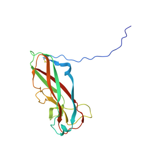
High-resolution cryo-EM analysis visualizes hydrated type I and IV pilus structures from enterotoxigenic Escherichia coli.
Kawahara, K., Oki, H., Iimori, M., Muramoto, R., Imai, T., Gerle, C., Shigematsu, H., Matsuda, S., Iida, T., Nakamura, S.(2025) Structure 33: 1040
- PubMed: 40220752 Search on PubMed
- DOI: https://doi.org/10.1016/j.str.2025.03.010
- Primary Citation Related Structures:
9IUF, 9IUG - PubMed Abstract:
Pathogenic bacteria utilize a variety of pilus filaments to colonize intestinal epithelia, including those synthesized by the chaperone-usher or type IV pilus assembly pathway. Despite the importance of these filaments as potential drug and vaccine targets, their large size and dynamic nature make high-resolution structure determination challenging. Here, we used cryo-electron microscopy (cryo-EM) and whole-genome sequencing to determine the structures of type I and IV pili expressed in enterotoxigenic Escherichia coli. Well-defined cryo-EM maps at resolutions of 2.2 and 1.8 Å for type I and IV pilus, respectively, facilitated the de novo structural modeling for these filaments, revealing side-chain structures in detail. We resolved thousands of hydrated water molecules around and within the inner core of the filaments, which stabilize the otherwise metastable quaternary subunit assembly. The high-resolution structures offer novel insights into subunit-subunit interactions, and provide important clues to understand pilus assembly, stability, and flexibility.
- Graduate School of Pharmaceutical Sciences, Osaka University, Suita, Osaka 565-0871, Japan; Center for Infectious Disease Education and Research, Osaka University, Suita, Osaka 565-0871, Japan. Electronic address: kkkazuki@phs.osaka-u.ac.jp.
Organizational Affiliation: