Assembly and breakage of head-to-head double hexamer reveals mpox virus E5-catalyzed DNA unwinding initiation.
Cheng, Y., Han, P., Peng, Q., Liu, R., Liu, H., Yuan, B., Zhao, Y., Kuai, L., Qi, J., Miao, K., Shi, Y., Gao, G.F., Wang, H.(2025) Nat Commun 16: 5176-5176
- PubMed: 40467608 Search on PubMedSearch on PubMed Central
- DOI: https://doi.org/10.1038/s41467-025-60539-1
- Primary Citation Related Structures:
9ILY, 9ILZ, 9IM0, 9IM1, 9IM2, 9IM3 - PubMed Abstract:
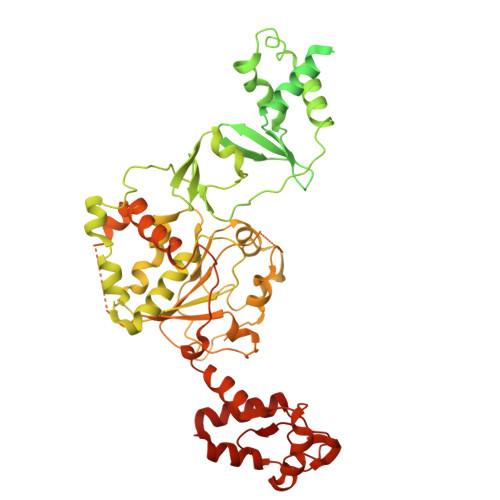
The replicative helicase-catalyzed unwinding of the DNA double helix is the initiation of DNA replication. Helicases and primases are functionally related enzymes that have even been expressed as fusion proteins in some organisms and viruses. However, the mechanism underlying DNA unwinding initiation by these helicase-primase fusion enzymes and the functional association between domains have not been elucidated. Herein, we report the cryo-EM structures of mpox virus E5, the founding member of these helicase-primase enzymes, in various enzymatic stages. Notably, E5 forms a head-to-head double hexamer encircling dsDNA, disrupted by the conformational rearrangement of primase domains upon nucleotide incorporation. Five E5-ssDNA-ATP structures further support an ATP cycle-driven non-classical escort model for E5 translocation. Finally, the helicase domain is found to enhance the primase function as a DNA scaffold. Together, our data shed light on the E5-mediated DNA unwinding model including dsDNA loading, DNA melting, ssDNA translocation, and provide a reasonable interpretation for evolutionary preservation of helicase-primase fusion from a functional perspective.
- College of Veterinary Medicine, Henan Agricultural University, Zhengzhou, Henan, China.
Organizational Affiliation: