9II2 | pdb_00009ii2

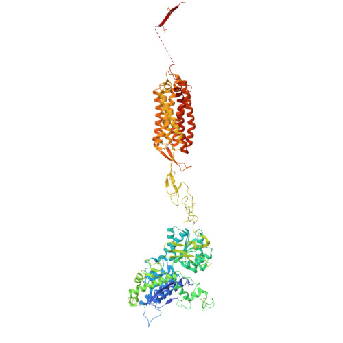
Cryo-EM Structure of the 2:2 Complex of mGlu3 and beta-arrestin1


Experimental Data Snapshot

  • Method: ELECTRON MICROSCOPY
  • Resolution: 3.70 Å
  • Aggregation State: PARTICLE 
  • Reconstruction Method: SINGLE PARTICLE 

wwPDB Validation 3D Report Full Report

Validation slider image for 9II2

This is version 1.3 of the entry. See complete history. 

Literature

Molecular basis of beta-arrestin coupling to the metabotropic glutamate receptor mGlu3.

Wen, T., Du, M., Lu, Y., Jia, N., Lu, X., Liu, N., Chang, S., Zhang, X., Shen, Y., Yang, X.

(2025) Nat Chem Biol 21: 1262-1269

  • DOI: https://doi.org/10.1038/s41589-025-01858-8
  • Primary Citation Related Structures: 
    9II2, 9II3

  • PubMed Abstract: 

    β-Arrestins (βarrs) mediate the desensitization and internalization of activated G-protein-coupled receptors (GPCRs). The molecular mechanism by which dimeric family C GPCR members recruit arrestins remains elusive. Here we report two structures of metabotropic glutamate receptor subtype 3 (mGlu3) coupled to βarr1, with stoichiometries of 2:1 and 2:2. The L-glutamate-bound mGlu3 dimer adopts an inactive state, with both Venus flytrap domains closed, engaging βarr1 either asymmetrically or symmetrically. The transmembrane domain of the mGlu3 protomer interacts with βarr1 through a binding pocket formed by three intracellular loops and an ordered C-terminal region. Three phosphorylation sites (pS857, pS859 and pT860) on the C-terminal tail of mGlu3 engage the N domain of βarr1. βarr1 stabilizes mGlu3 in an inactive conformation, characterized by a TM3/TM4-TM3/TM4 dimeric interface, previously observed in the negative allosteric modulator-bound structure of mGlu3. Our findings provide important insights into βarr-mediated inactivation of family C GPCRs.


  • Organizational Affiliation: 
    • State Key Laboratory of Medicinal Chemical Biology and Frontiers Science Center for Cell Responses, College of Life Sciences, Nankai University, Tianjin, China.

Macromolecule Content 

  • Total Structure Weight: 350.17 kDa 
  • Atom Count: 21,112 
  • Modeled Residue Count: 2,748 
  • Deposited Residue Count: 3,096 
  • Unique protein chains: 3

Macromolecules

Find similar proteins by:|  3D Structure
Entity ID: 1
MoleculeChains  Sequence LengthOrganismDetailsImage
Metabotropic glutamate receptor 3A [auth R],
D [auth B]
879Homo sapiensMutation(s): 0 
Gene Names: GRM3, GPRC1C, MGLUR3
Membrane Entity: Yes 
UniProt & NIH Common Fund Data Resources
Find proteins for Q14832 (Homo sapiens)
Explore Q14832 
Go to UniProtKB:  Q14832
PHAROS:  Q14832
GTEx:  ENSG00000198822 
Entity Groups
Sequence Clusters30% Identity50% Identity70% Identity90% Identity95% Identity100% Identity
UniProt GroupQ14832
Glycosylation
Glycosylation Sites: 1Go to GlyGen: Q14832-1
Sequence Annotations
Expand
Reference Sequence
Find similar proteins by:|  3D Structure
Entity ID: 2
MoleculeChains  Sequence LengthOrganismDetailsImage
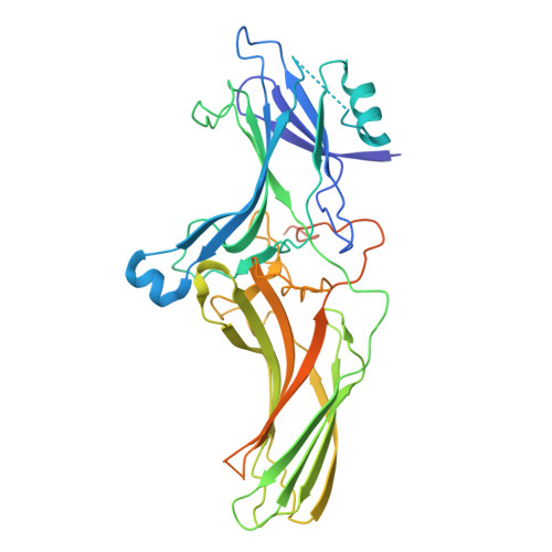
Beta-arrestin-1B [auth A],
E [auth C]
418Homo sapiensMutation(s): 0 
Gene Names: ARRB1, ARR1
Membrane Entity: Yes 
UniProt & NIH Common Fund Data Resources
Find proteins for P49407 (Homo sapiens)
Explore P49407 
Go to UniProtKB:  P49407
PHAROS:  P49407
GTEx:  ENSG00000137486 
Entity Groups
Sequence Clusters30% Identity50% Identity70% Identity90% Identity95% Identity100% Identity
UniProt GroupP49407
Sequence Annotations
Expand
Reference Sequence
Find similar proteins by:|  3D Structure
Entity ID: 3
MoleculeChains  Sequence LengthOrganismDetailsImage
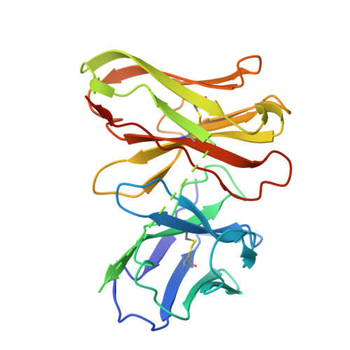
scFv30C [auth S],
F [auth D]
251Mus musculusMutation(s): 0 
Entity Groups
Sequence Clusters30% Identity50% Identity70% Identity90% Identity95% Identity100% Identity
Sequence Annotations
Expand
Reference Sequence

Oligosaccharides

Help  
Entity ID: 4
MoleculeChains Length2D Diagram GlycosylationD Interactions
2-acetamido-2-deoxy-beta-D-glucopyranose-(1-4)-2-acetamido-2-deoxy-beta-D-glucopyranoseG [auth E],
H [auth F]
2N-Glycosylation
Glycosylation Resources
GlyTouCan: G42666HT
GlyCosmos: G42666HT
GlyGen: G42666HT

Small Molecules

Ligands 2 Unique
IDChains Name / Formula / InChI Key2D Diagram3D Interactions
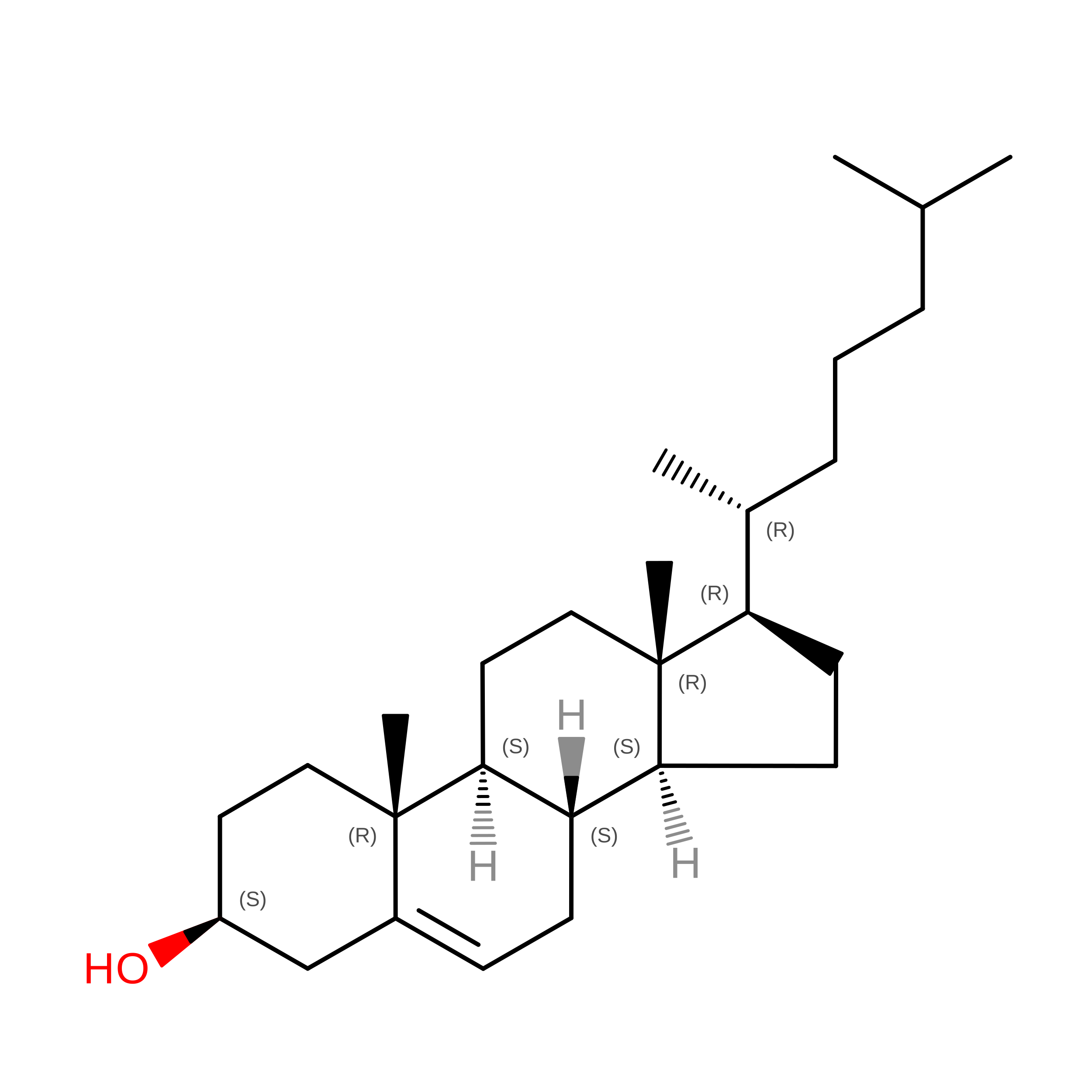
CLR

Query on CLR


Download:Ideal Coordinates CCD File
J [auth R]
K [auth R]
L [auth R]
N [auth B]
O [auth B]
J [auth R],
K [auth R],
L [auth R],
N [auth B],
O [auth B],
P [auth B]
CHOLESTEROL
C27 H46 O
HVYWMOMLDIMFJA-DPAQBDIFSA-N
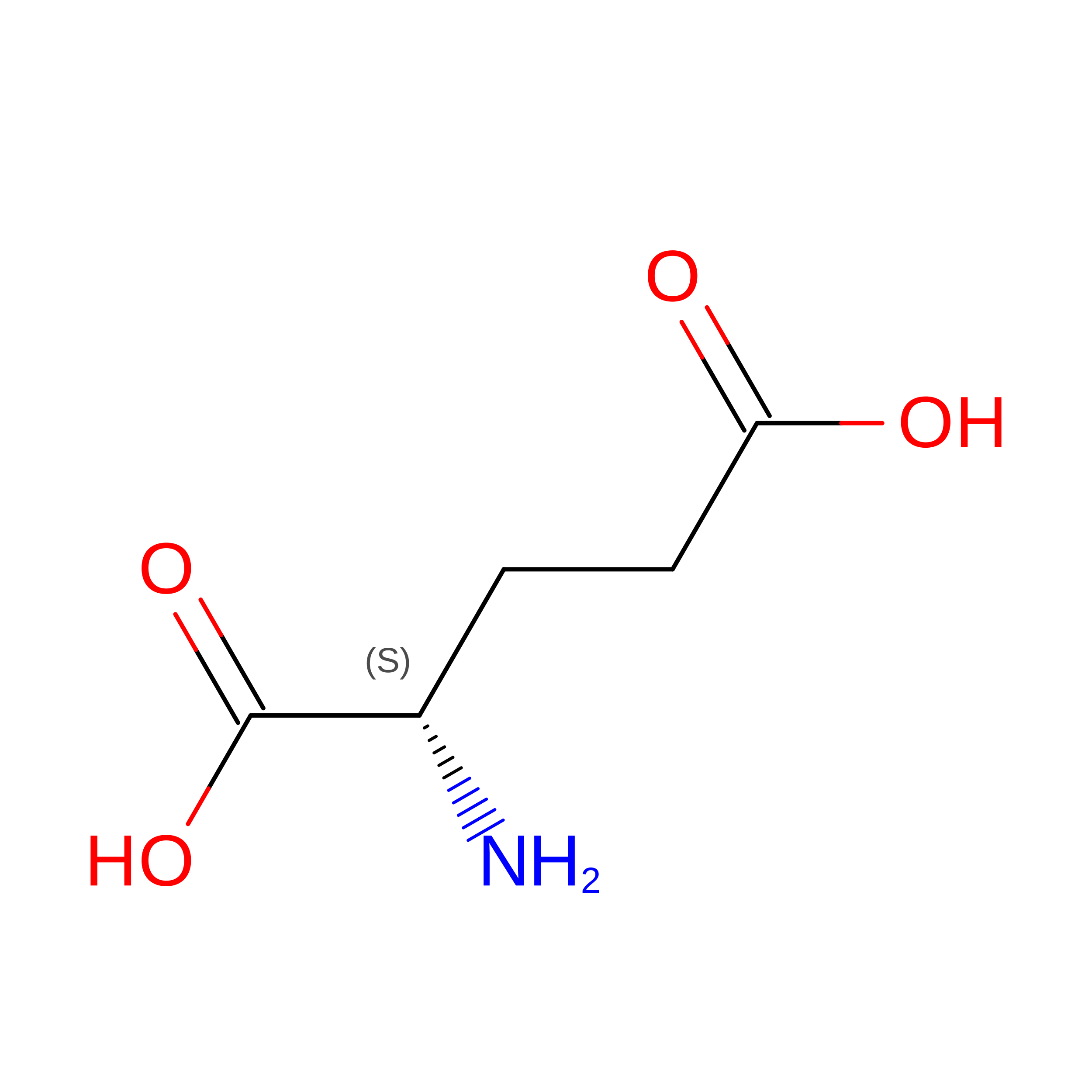
GLU

Query on GLU


Download:Ideal Coordinates CCD File
I [auth R],
M [auth B]
GLUTAMIC ACID
C5 H9 N O4
WHUUTDBJXJRKMK-VKHMYHEASA-N
Modified Residues  2 Unique
IDChains TypeFormula2D DiagramParent
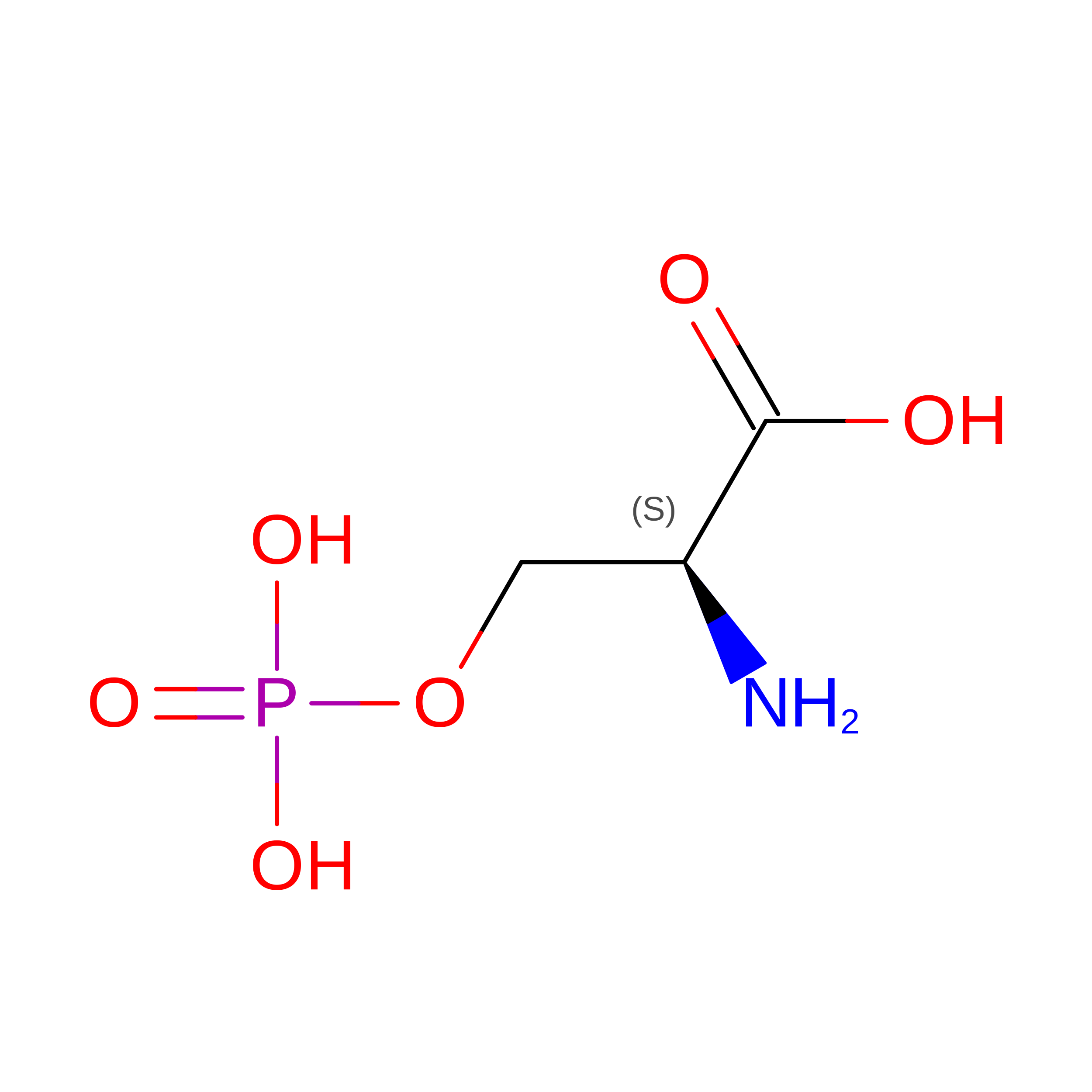
SEP
Query on SEP
A [auth R],
D [auth B]
L-PEPTIDE LINKINGC3 H8 N O6 PSER
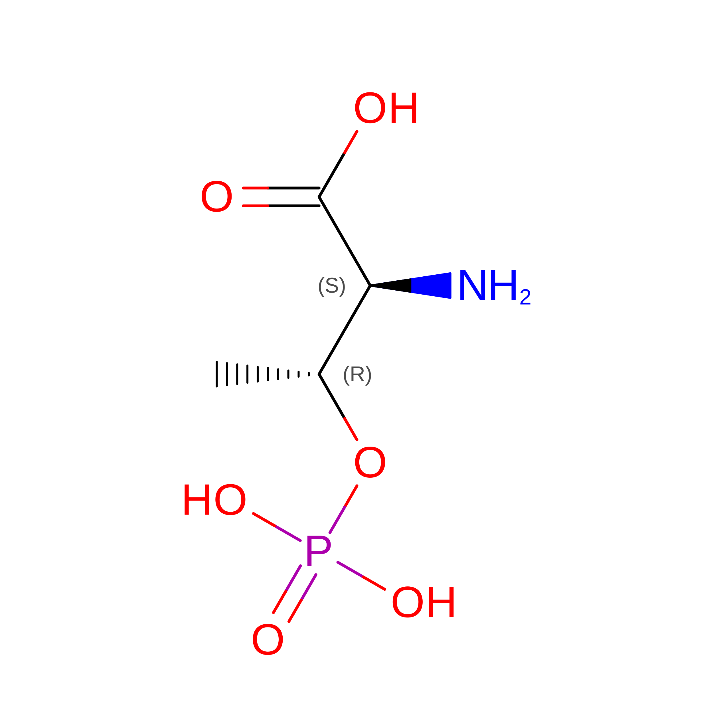
TPO
Query on TPO
A [auth R],
D [auth B]
L-PEPTIDE LINKINGC4 H10 N O6 PTHR

Experimental Data & Validation

Experimental Data

  • Method: ELECTRON MICROSCOPY
  • Resolution: 3.70 Å
  • Aggregation State: PARTICLE 
  • Reconstruction Method: SINGLE PARTICLE 
EM Software:
TaskSoftware PackageVersion
MODEL REFINEMENTPHENIX

Structure Validation

View Full Validation Report


Entry History 

& Funding Information

Deposition Data


Funding OrganizationLocationGrant Number
National Natural Science Foundation of China (NSFC)China--

Revision History  (Full details and data files)

  • Version 1.0: 2025-03-05
    Type: Initial release
  • Version 1.1: 2025-03-19
    Changes: Data collection, Database references
  • Version 1.2: 2025-07-23
    Changes: Data collection
  • Version 1.3: 2025-08-13
    Changes: Data collection, Database references