9IGO | pdb_00009igo

PR3 S203A I221N W222N G223T mutant in complex with the extracellular domain of CD177


Experimental Data Snapshot

  • Method: X-RAY DIFFRACTION
  • Resolution: 1.50 Å
  • R-Value Free: 
    0.184 (Depositor), 0.184 (DCC) 
  • R-Value Work: 
    0.146 (Depositor), 0.146 (DCC) 
  • R-Value Observed: 
    0.148 (Depositor) 

Starting Models: experimental
View more details

wwPDB Validation   3D Report Full Report


Ligand Structure Quality Assessment 


This is version 1.1 of the entry. See complete history


Literature

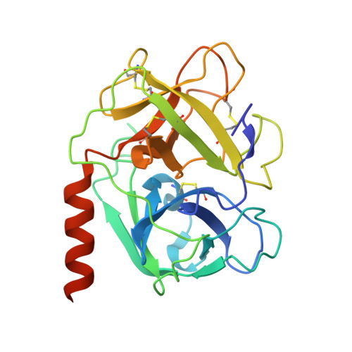
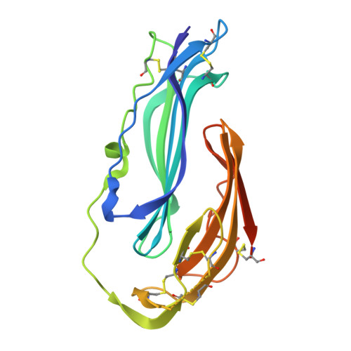
Structures of proteinase 3 and the CD177 receptor complex reveal a major autoantibody epitope.

Zheng-Gerard, C.Joha, J.Carrasquero, M.El Omari, K.Lowe, E.Dubey, S.Draper, S.J.Chang, Y.C.Lin, H.H.Salama, A.D.McHugh, K.Seiradake, E.

(2026) EMBO Rep 

  • DOI: https://doi.org/10.1038/s44319-026-00716-5
  • Primary Citation of Related Structures:  
    9IGO, 9IGP

  • PubMed Abstract: 

    Granulomatosis with polyangiitis is a life-threatening systemic vasculitis, characterised by anti-neutrophil cytoplasmic autoantibodies (ANCA) most commonly against proteinase 3 (PR3), a protease expressed intracellularly and on the surface of neutrophils. Most cell surface PR3 is bound to the receptor CD177; however, the molecular mechanism of the interactions is not well understood. Here, we present crystal structures of CD177 in complex with PR3 and unliganded CD177. We describe a mainly hydrophobic binding interface between PR3 and CD177, involving the first two Ly6/uPAR (LU) domains of CD177. These form a globular structure which is connected to downstream domains via a flexible linker. Using a panel of PR3-ANCA-positive patient samples, we show that a significant proportion of ANCAs target the CD177-binding site of PR3 in these samples. Structure-guided mutation of the CD177-binding site on PR3 is effective in reducing PR3-ANCA binding. The results demonstrate that the CD177-binding surface of PR3 harbours a major PR3-ANCA epitope, and that the extent of binding to this surface varies between different patients.


  • Organizational Affiliation
    • Department of Biochemistry, University of Oxford, South Parks Road, Oxford, OX1 3QU, UK.

Macromolecules
Find similar proteins by:  (by identity cutoff)  |  3D Structure
Entity ID: 1
MoleculeChains Sequence LengthOrganismDetailsImage
Myeloblastin238Homo sapiensMutation(s): 5 
Gene Names: PRTN3MBN
EC: 3.4.21.76
UniProt & NIH Common Fund Data Resources
Find proteins for P24158 (Homo sapiens)
Explore P24158 
Go to UniProtKB:  P24158
PHAROS:  P24158
GTEx:  ENSG00000196415 
Entity Groups  
Sequence Clusters30% Identity50% Identity70% Identity90% Identity95% Identity100% Identity
UniProt GroupP24158
Glycosylation
Glycosylation Sites: 3Go to GlyGen: P24158-1
Sequence Annotations
Expand
  • Reference Sequence
Find similar proteins by:  (by identity cutoff)  |  3D Structure
Entity ID: 2
MoleculeChains Sequence LengthOrganismDetailsImage
CD177 antigen194Homo sapiensMutation(s): 0 
Gene Names: CD177NB1PRV1UNQ595/PRO1181
UniProt & NIH Common Fund Data Resources
Find proteins for Q8N6Q3 (Homo sapiens)
Explore Q8N6Q3 
Go to UniProtKB:  Q8N6Q3
PHAROS:  Q8N6Q3
GTEx:  ENSG00000204936 
Entity Groups  
Sequence Clusters30% Identity50% Identity70% Identity90% Identity95% Identity100% Identity
UniProt GroupQ8N6Q3
Glycosylation
Glycosylation Sites: 1Go to GlyGen: Q8N6Q3-1
Sequence Annotations
Expand
  • Reference Sequence
Oligosaccharides

Help

Entity ID: 3
MoleculeChains Length2D Diagram Glycosylation3D Interactions
2-acetamido-2-deoxy-beta-D-glucopyranose-(1-4)-2-acetamido-2-deoxy-beta-D-glucopyranose
C, D
2N-Glycosylation
Glycosylation Resources
GlyTouCan:  G42666HT
GlyCosmos:  G42666HT
GlyGen:  G42666HT
Small Molecules
Ligands 4 Unique
IDChains Name / Formula / InChI Key2D Diagram3D Interactions
NAG
Query on NAG

Download Ideal Coordinates CCD File 
E [auth A],
F [auth A]
2-acetamido-2-deoxy-beta-D-glucopyranose
C8 H15 N O6
OVRNDRQMDRJTHS-FMDGEEDCSA-N
GOL
Query on GOL

Download Ideal Coordinates CCD File 
H [auth A],
L [auth B]
GLYCEROL
C3 H8 O3
PEDCQBHIVMGVHV-UHFFFAOYSA-N
ACT
Query on ACT

Download Ideal Coordinates CCD File 
I [auth A],
J [auth A],
K [auth A]
ACETATE ION
C2 H3 O2
QTBSBXVTEAMEQO-UHFFFAOYSA-M
CL
Query on CL

Download Ideal Coordinates CCD File 
G [auth A]CHLORIDE ION
Cl
VEXZGXHMUGYJMC-UHFFFAOYSA-M
Experimental Data & Validation

Experimental Data

  • Method: X-RAY DIFFRACTION
  • Resolution: 1.50 Å
  • R-Value Free:  0.184 (Depositor), 0.184 (DCC) 
  • R-Value Work:  0.146 (Depositor), 0.146 (DCC) 
  • R-Value Observed: 0.148 (Depositor) 
Space Group: P 1 21 1
Unit Cell:
Length ( Å )Angle ( ˚ )
a = 39.76α = 90
b = 120.735β = 96.99
c = 61.983γ = 90
Software Package:
Software NamePurpose
PHENIXrefinement
DIALSdata reduction
Aimlessdata scaling
PHASERphasing

Structure Validation

View Full Validation Report


Ligand Structure Quality Assessment 


Entry History & Funding Information

Deposition Data


Funding OrganizationLocationGrant Number
Wellcome TrustUnited Kingdom202827/Z/16/Z

Revision History  (Full details and data files)

  • Version 1.0: 2026-02-25
    Type: Initial release
  • Version 1.1: 2026-03-04
    Changes: Database references