


Best-in-Class FSP1 Inhibitors Unraveled By AI-Enhanced Adaptive Screening Platform
Cecchini, D., Mattevi, A.To be published.
Experimental Data Snapshot
Starting Model: experimental
View more details
Entity ID: 1 | |||||
|---|---|---|---|---|---|
| Molecule | Chains | Sequence Length | Organism | Details | Image |
| FSP1 (tetrapod ancestor) | 368 | Mammalia | Mutation(s): 0 EC: 1.6.5 | 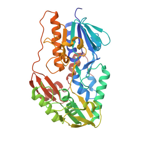 | |
UniProt | |||||
Entity Groups | |||||
| Sequence Clusters | 30% Identity50% Identity70% Identity90% Identity95% Identity100% Identity | ||||
| UniProt Group | E1BR24 | ||||
Sequence AnnotationsExpand | |||||
Reference Sequence | |||||
| Ligands 7 Unique | |||||
|---|---|---|---|---|---|
| ID | Chains | Name / Formula / InChI Key | 2D Diagram | 3D Interactions | |
| FAD Download:Ideal Coordinates CCD File | C [auth A], J [auth B] | FLAVIN-ADENINE DINUCLEOTIDE C27 H33 N9 O15 P2 VWWQXMAJTJZDQX-UYBVJOGSSA-N |  | ||
| NAD Download:Ideal Coordinates CCD File | D [auth A], K [auth B] | NICOTINAMIDE-ADENINE-DINUCLEOTIDE C21 H27 N7 O14 P2 BAWFJGJZGIEFAR-NNYOXOHSSA-N |  | ||
| A1I34 (Subject of Investigation/LOI) Download:Ideal Coordinates CCD File | E [auth A], L [auth B] | ~{N}-[3-[4-(5-phenyl-4~{H}-1,2,4-triazol-3-yl)piperidin-1-yl]carbonylphenyl]benzamide C27 H25 N5 O2 PMGKFAXGJYRVER-UHFFFAOYSA-N |  | ||
| PEG Download:Ideal Coordinates CCD File | F [auth A] | DI(HYDROXYETHYL)ETHER C4 H10 O3 MTHSVFCYNBDYFN-UHFFFAOYSA-N |  | ||
| CA Download:Ideal Coordinates CCD File | G [auth A] | CALCIUM ION Ca BHPQYMZQTOCNFJ-UHFFFAOYSA-N |  | ||
| CL Download:Ideal Coordinates CCD File | H [auth A] | CHLORIDE ION Cl VEXZGXHMUGYJMC-UHFFFAOYSA-M |  | ||
| MG Download:Ideal Coordinates CCD File | I [auth A] | MAGNESIUM ION Mg JLVVSXFLKOJNIY-UHFFFAOYSA-N |  | ||
| Length ( Å ) | Angle ( ˚ ) |
|---|---|
| a = 43.85 | α = 90 |
| b = 79.335 | β = 99.08 |
| c = 113.373 | γ = 90 |
| Software Name | Purpose |
|---|---|
| REFMAC | refinement |
| Aimless | data scaling |
| XDS | data reduction |
| PHASER | phasing |
| Funding Organization | Location | Grant Number |
|---|---|---|
| European Research Council (ERC) | European Union | 101094471 |
| Italian Association for Cancer Research | Italy | 28754 |


