9I91 | pdb_00009i91

Ku from Mycobacterium tuberculosis bound to DNA


Experimental Data Snapshot

  • Method: ELECTRON MICROSCOPY
  • Resolution: 2.96 Å
  • Aggregation State: FILAMENT 
  • Reconstruction Method: SINGLE PARTICLE 

wwPDB Validation   3D Report Full Report


This is version 1.0 of the entry. See complete history


Literature

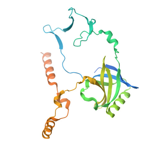
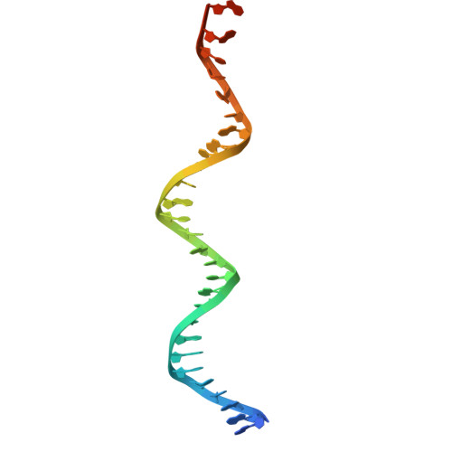
Oligomerisation of Ku from Mycobacterium tuberculosis promotes DNA synapsis.

Zahid, S.Baconnais, S.Smith, H.Atwal, S.Bates, L.Read, H.Chadda, A.Morati, F.Bedwell, T.Stender, E.G.P.Walter, J.Hardwick, S.W.Westerlund, F.Galburt, E.Le Cam, E.Pyne, A.Mukamolova, G.V.Chaplin, A.K.

(2025) Nat Commun 16: 10568-10568

  • DOI: https://doi.org/10.1038/s41467-025-65609-y
  • Primary Citation of Related Structures:  
    9I91

  • PubMed Abstract: 

    Mycobacterium tuberculosis (Mtb), the causative agent of tuberculosis (TB), is estimated to infect nearly one-quarter of the global population. A key factor in its resilience and persistence is its robust DNA repair capacity. Non-homologous end joining (NHEJ) is the primary pathway for repairing DNA double-strand breaks (DSBs) in many organisms, including Mtb, where it is mediated by the Ku protein and the multifunctional LigD enzyme. In this study, we demonstrate that Ku is essential for mycobacterial survival under DNA-damaging conditions. Using cryogenic electron microscopy (cryo-EM), we solved high-resolution structures of both the apo and DNA-bound forms of the Ku-Mtb homodimer. Our structural and biophysical analyses reveal that Ku forms an extended proteo-filament upon binding DNA. We identify critical residues involved in filament formation and DNA synapsis and show that their mutation severely impairs bacterial viability. Furthermore, we propose a model in which the C-terminus of Ku regulates DNA binding and loading and facilitates subsequent recruitment of LigD. These findings provide unique insights into bacterial DNA repair and guide future therapeutics.


  • Organizational Affiliation
    • Leicester Institute for Structural and Chemical Biology, Department of Molecular and Cell Biology, University of Leicester, Leicester, UK.

Macromolecules

Find similar proteins by:  (by identity cutoff)  |  3D Structure
Entity ID: 1
MoleculeChains Sequence LengthOrganismDetailsImage
Non-homologous end joining protein Ku
A, B, C, D, E
A, B, C, D, E, F
296Mycobacterium tuberculosisMutation(s): 0 
Gene Names: mkuRv0937c
UniProt
Find proteins for P9WKD9 (Mycobacterium tuberculosis (strain ATCC 25618 / H37Rv))
Explore P9WKD9 
Go to UniProtKB:  P9WKD9
Entity Groups  
Sequence Clusters30% Identity50% Identity70% Identity90% Identity95% Identity100% Identity
UniProt GroupP9WKD9
Sequence Annotations
Expand
  • Reference Sequence
Find similar nucleic acids by:  (by identity cutoff)  |  3D Structure
Entity ID: 2
MoleculeChains LengthOrganismImage
DNA 1
G, I
32DNA molecule
Sequence Annotations
Expand
  • Reference Sequence
Find similar nucleic acids by:  (by identity cutoff)  |  3D Structure
Entity ID: 3
MoleculeChains LengthOrganismImage
DNA 2
H, J
32DNA molecule
Sequence Annotations
Expand
  • Reference Sequence
Experimental Data & Validation

Experimental Data

  • Method: ELECTRON MICROSCOPY
  • Resolution: 2.96 Å
  • Aggregation State: FILAMENT 
  • Reconstruction Method: SINGLE PARTICLE 
EM Software:
TaskSoftware PackageVersion
MODEL REFINEMENTPHENIX

Structure Validation

View Full Validation Report


Entry History & Funding Information

Deposition Data


Funding OrganizationLocationGrant Number
Medical Research Council (MRC, United Kingdom)United KingdomMR/X00029X/1
The Lister Institute of Preventive MedicineUnited Kingdom--

Revision History  (Full details and data files)

  • Version 1.0: 2025-12-10
    Type: Initial release