9HZK | pdb_00009hzk

sdAb9 in complex with PfCSP aTSR domain


Experimental Data Snapshot

  • Method: X-RAY DIFFRACTION
  • Resolution: 2.07 Å
  • R-Value Free: 
    0.226 (Depositor), 0.224 (DCC) 
  • R-Value Work: 
    0.184 (Depositor), 0.184 (DCC) 
  • R-Value Observed: 
    0.186 (Depositor) 

Starting Models: experimental, in silico
View more details

wwPDB Validation 3D Report Full Report

Validation slider image for 9HZK

This is version 1.3 of the entry. See complete history

Literature

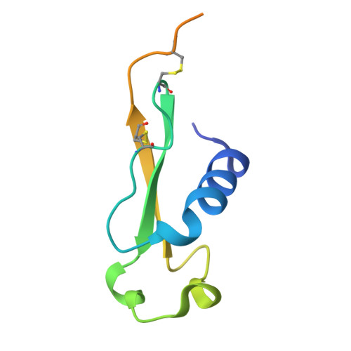
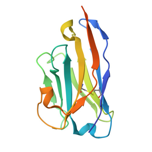
Evidence for a model of conformational change by the Plasmodium falciparum circumsporozoite protein during sporozoite development in the mosquito host through the use of camelid single-domain antibodies.

Geens, R.De Vocht, L.Aguirre-Botero, M.C.Vincke, C.Romao, E.Magez, S.Muyldermans, S.Amino, R.Sterckx, Y.G.-.J.

(2025) Infect Immun 93: e0008125-e0008125

  • DOI: https://doi.org/10.1128/iai.00081-25
  • Primary Citation Related Structures: 
    9HZJ, 9HZK

  • PubMed Abstract: 

    Plasmodium sporozoites (SPZs) are formed in the Anopheles mosquito midgut from where they travel to the salivary glands and subsequently to the mammalian liver after deposition into the skin. The SPZ's main surface antigen, the circumsporozoite protein (CSP), plays a pivotal role in SPZ biology and constitutes the immunodominant target for host antibodies. In this study, we raised single-domain antibodies (sdAbs) against CSP from P. falciparum (PfCSP) by immunizing two alpacas with recombinant versions of the antigen. We found that all identified sdAbs specifically target PfCSP's globular [Formula: see text]TSR domain without cross-reacting with P. berghei CSP. Further characterization revealed that most sdAbs recognize native PfCSP on the SPZ surface, although they do not have any inhibitory effect on hepatocyte binding and invasion. Structural studies showed that all binders target the previously identified [Formula: see text]-epitope, confirming the non-protective nature of this epitope. Comparison of sdAb binding to midgut and salivary gland SPZs revealed a shift in the exposure and accessibility of the [Formula: see text]-epitope. Hence, our findings provide further evidence that CSP undergoes structural changes during SPZ development in the mosquito host.


  • Organizational Affiliation
    • Laboratory of Medical Biochemistry (LMB) and the Infla-Med Centre of Excellence, Department of Pharmaceutical Sciences, University of Antwerp, Antwerp, Belgium.

Macromolecule Content 

  • Total Structure Weight: 27.46 kDa 
  • Atom Count: 1,641 
  • Modeled Residue Count: 192 
  • Deposited Residue Count: 235 
  • Unique protein chains: 2

Macromolecules

Find similar proteins by:|  3D Structure
Entity ID: 1
MoleculeChains  Sequence LengthOrganismDetailsImage
Circumsporozoite proteinA [auth B]91Plasmodium falciparum NF54Mutation(s): 0 
Gene Names: CSP
UniProt
Find proteins for P19597 (Plasmodium falciparum (isolate NF54))
Explore P19597 
Go to UniProtKB:  P19597
Entity Groups
Sequence Clusters30% Identity50% Identity70% Identity90% Identity95% Identity100% Identity
UniProt GroupP19597
Sequence Annotations
Expand
Reference Sequence
Find similar proteins by:|  3D Structure
Entity ID: 2
MoleculeChains  Sequence LengthOrganismDetailsImage
sdAb9B [auth A]144Lama glamaMutation(s): 0 

Small Molecules

Ligands 2 Unique
IDChains Name / Formula / InChI Key2D Diagram3D Interactions
PEG

Query on PEG


Download:Ideal Coordinates CCD File
H [auth A],
J [auth A],
N [auth A]
DI(HYDROXYETHYL)ETHER
C4 H10 O3
MTHSVFCYNBDYFN-UHFFFAOYSA-N
GOL

Query on GOL


Download:Ideal Coordinates CCD File
C [auth B]
D [auth B]
E [auth B]
F [auth B]
G [auth B]
C [auth B],
D [auth B],
E [auth B],
F [auth B],
G [auth B],
I [auth A],
K [auth A],
L [auth A],
M [auth A],
O [auth A]
GLYCEROL
C3 H8 O3
PEDCQBHIVMGVHV-UHFFFAOYSA-N

Experimental Data & Validation

Experimental Data

  • Method: X-RAY DIFFRACTION
  • Resolution: 2.07 Å
  • R-Value Free:  0.226 (Depositor), 0.224 (DCC) 
  • R-Value Work:  0.184 (Depositor), 0.184 (DCC) 
  • R-Value Observed: 0.186 (Depositor) 
Space Group: P 32
Unit Cell:
Length ( Å )Angle ( ˚ )
a = 53.74α = 90
b = 53.74β = 90
c = 60.84γ = 120
Software Package:
Software NamePurpose
PHENIXrefinement
XDSdata reduction
XDSdata scaling
PHASERphasing

Structure Validation

View Full Validation Report


Entry History 

& Funding Information

Deposition Data


Funding OrganizationLocationGrant Number
Other governmentBelgiumUAntwerp BOF DOCPRO4-NIEUWZAP 40043
Other governmentBelgiumUAntwerp BOF infrastructure 41391
Research Foundation - Flanders (FWO)Belgium11P4B24N

Revision History  (Full details and data files)

  • Version 1.0: 2025-04-23
    Type: Initial release
  • Version 1.1: 2025-05-07
    Changes: Database references
  • Version 1.2: 2025-06-18
    Changes: Database references
  • Version 1.3: 2025-06-25
    Changes: Database references