9HVM | pdb_00009hvm

In-cell Structure of Pyrenoid Rubisco


Experimental Data Snapshot

  • Method: ELECTRON MICROSCOPY
  • Resolution: 8.10 Å
  • Aggregation State: CELL 
  • Reconstruction Method: SUBTOMOGRAM AVERAGING 

Starting Models: experimental
View more details

wwPDB Validation 3D Report Full Report

Validation slider image for 9HVM

This is version 1.1 of the entry. See complete history

Literature

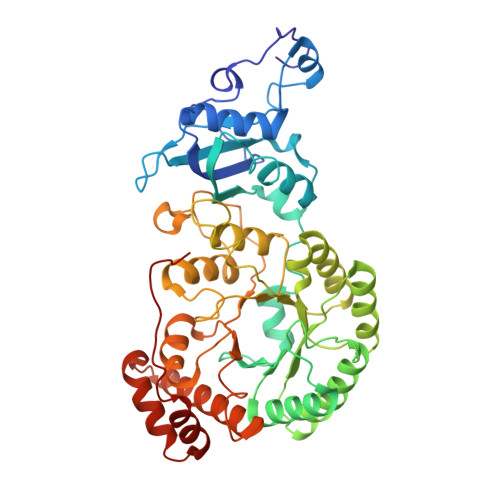
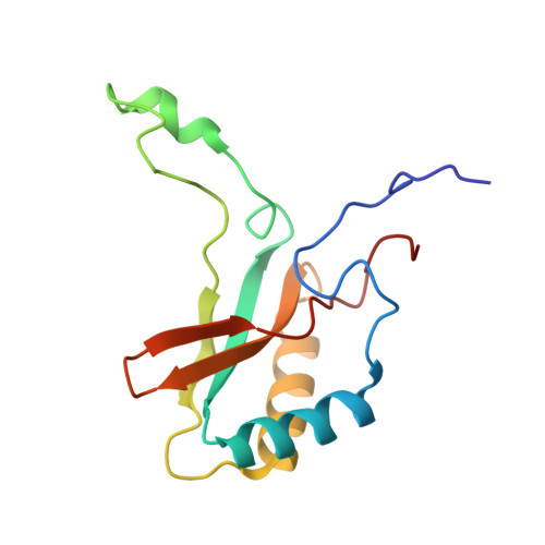
In-cell structure and variability of pyrenoid Rubisco.

Elad, N.Hou, Z.Dumoux, M.Ramezani, A.Perilla, J.R.Zhang, P.

(2025) Nat Commun 16: 7763-7763

  • DOI: https://doi.org/10.1038/s41467-025-62998-y
  • Primary Citation Related Structures: 
    9HVM

  • PubMed Abstract: 

    Ribulose-1,5-bisphosphate carboxylase/oxygenase (Rubisco) is central to global CO 2 fixation. In eukaryotic algae, its catalytic efficiency is enhanced through the pyrenoid - a protein-dense organelle within the chloroplast that concentrates CO 2 . Although Rubisco structure has been extensively studied in vitro, its native structure, dynamics and interactions within the pyrenoid remain elusive. Here, we present the native Rubisco structure inside the green alga Chlamydomonas reinhardtii determined by cryo-electron tomography and subtomogram averaging of cryo-focused ion beam milled cells. Multiple structural subsets of Rubisco are identified, stochastically distributed throughout the pyrenoid. While Rubisco adopts an active conformation in the best-resolved map, comparison among the subsets reveals significant local variations at the active site, at the large subunit dimer interfaces, and at binding protein contact regions. These findings offer a comprehensive understanding of the structure, dynamics, and functional organization of native Rubisco within the pyrenoid, providing valuable insights into its critical role in CO 2 fixation.


  • Organizational Affiliation
    • Department of Chemical Research Support, Weizmann Institute of Science, Rehovot, Israel.

Macromolecule Content 

  • Total Structure Weight: 538.49 kDa 
  • Atom Count: 37,816 
  • Modeled Residue Count: 4,808 
  • Deposited Residue Count: 4,808 
  • Unique protein chains: 2

Macromolecules

Find similar proteins by:|  3D Structure
Entity ID: 1
MoleculeChains  Sequence LengthOrganismDetailsImage
Ribulose bisphosphate carboxylase large chain
A, C, E, G, I
A, C, E, G, I, K, M, O
469Chlamydomonas reinhardtiiMutation(s): 0 
EC: 4.1.1.39
UniProt
Find proteins for P00877 (Chlamydomonas reinhardtii)
Explore P00877 
Go to UniProtKB:  P00877
Entity Groups
Sequence Clusters30% Identity50% Identity70% Identity90% Identity95% Identity100% Identity
UniProt GroupP00877
Sequence Annotations
Expand
Reference Sequence
Find similar proteins by:|  3D Structure
Entity ID: 2
MoleculeChains  Sequence LengthOrganismDetailsImage
Ribulose bisphosphate carboxylase small subunit, chloroplastic 1
B, D, F, H, J
B, D, F, H, J, L, N, P
132Chlamydomonas reinhardtiiMutation(s): 0 
Gene Names: RBCS1RBCS-1CHLRE_02g120100v5CHLREDRAFT_82986
UniProt
Find proteins for P00873 (Chlamydomonas reinhardtii)
Explore P00873 
Go to UniProtKB:  P00873
Entity Groups
Sequence Clusters30% Identity50% Identity70% Identity90% Identity95% Identity100% Identity
UniProt GroupP00873
Sequence Annotations
Expand
Reference Sequence

Experimental Data & Validation

Experimental Data

  • Method: ELECTRON MICROSCOPY
  • Resolution: 8.10 Å
  • Aggregation State: CELL 
  • Reconstruction Method: SUBTOMOGRAM AVERAGING 
EM Software:
TaskSoftware PackageVersion
RECONSTRUCTIONRELION4.0

Structure Validation

View Full Validation Report


Entry History 

& Funding Information

Deposition Data


Funding OrganizationLocationGrant Number
Biotechnology and Biological Sciences Research Council (BBSRC)United KingdomBB/S003339/1

Revision History  (Full details and data files)

  • Version 1.0: 2025-03-05
    Type: Initial release
  • Version 1.1: 2025-09-17
    Changes: Data collection, Database references