Yersinia ruckeri YRB periplasmic binding protein YiuA selectively recognizes a Fe(III)-mono-catecholate siderophore.
Thomsen, E., Thompson, S., Stow, P.R., Cukor, M., Grogan, G., Duhme-Klair, A.K., Butler, A.(2025) Chem Commun (Camb) 61: 17653-17656
- PubMed: 41098114 Search on PubMed
- DOI: https://doi.org/10.1039/d5cc05103g
- Primary Citation Related Structures:
9HRJ, 9HRP, 9HT5, 9IF3 - PubMed Abstract:
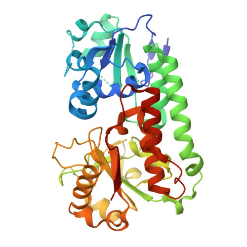
The marine pathogen Yersinia ruckeri synthesizes the tri-catecholate siderophore ruckerbactin, Rb, (DHB- L Arg- L Ser) 3 , to acquire iron during infection. Its biosynthetic gene cluster encodes a single periplasmic binding protein, RupB, which surprisingly does not bind Fe(III)-Rb nor the Fe(III) complexes of its hydrolysis products, the di- and mono-catecholate siderophores Rb DC and Rb MC , with biologically relevant affinities. Instead, the periplasmic binding protein YiuA, encoded in a different region of the chromosome, binds the 1 : 2 Fe(III) complex of the mono-catecholate Rb MC , Fe(III)-(Rb MC ) 2 . YiuA is the first periplasmic binding protein (PBP) to selectively recognize a mono-catecholate siderophore, the structural basis of which was illuminated through X-ray crystallography of YiuA bound to Fe(III)-(Rb MC ) 2 .
- University of California, Department of Chemistry & Biochemistry, Santa Barbara, CA 93106-9510, USA. butler@chem.ucsb.edu.
Organizational Affiliation: