9HR6 | pdb_00009hr6

cryoEM structure of amyloid fibrils formed by human RIPK1


Experimental Data Snapshot

  • Method: ELECTRON MICROSCOPY
  • Resolution: 2.57 Å
  • Aggregation State: HELICAL ARRAY 
  • Reconstruction Method: HELICAL 

Starting Model: in silico
View more details

wwPDB Validation 3D Report Full Report

Validation slider image for 9HR6

This is version 1.1 of the entry. See complete history

Literature

Structural basis for amyloid fibril assembly by the master cell-signaling regulator receptor-interacting protein kinase 1.

Polonio, P.Lopez-Alonso, J.P.Jiang, H.Andres-Campos, S.Escobedo-Gonzalez, F.C.Titaux-Delgado, G.A.Ubarretxena-Belandia, I.Mompean, M.

(2025) Nat Commun 16: 9607-9607

  • DOI: https://doi.org/10.1038/s41467-025-64621-6
  • Primary Citation Related Structures: 
    9HR6

  • PubMed Abstract: 

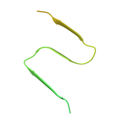
    Amyloid fibrils can form biologically relevant functional assemblies. The RIP homotypic interaction motifs (RHIMs) in receptor-interacting protein kinases 1 and 3 (RIPK1 and RIPK3) orchestrate the formation of amyloid-like fibrils essential for propagating cell death signals. While the structures of human RIPK3 (hRIPK3) homomeric fibrils and RIPK1-RIPK3 heteromeric fibrils have been elucidated, the atomic structure of human RIPK1 (hRIPK1) homomeric fibrils has remained elusive. We present a high-resolution structure of hRIPK1 RHIM-mediated amyloid fibrils, determined using an integrative approach combining cryoprobe-detected solid-state nuclear magnetic resonance spectroscopy and cryo-electron microscopy. The fibrils adopt an N-shaped fold consisting of three β-sheets stabilized by hydrophobic interactions and hydrogen bonding. A key hydrogen bond between N545 and G542 closes the β2-β3 loop, resulting in denser side-chain packing compared to hRIPK3 homomeric fibrils. These findings provide structural insights into how hRIPK1 homomeric fibrils nucleate hRIPK3 recruitment and fibrillization during necroptosis, offering broader perspectives on the molecular principles governing RHIM-mediated amyloid assembly and functional amyloids.


  • Organizational Affiliation
    • Instituto de Química Física Blas Cabrera (IQF-CSIC), Madrid, Spain.

Macromolecule Content 

  • Total Structure Weight: 48.28 kDa 
  • Atom Count: 930 
  • Modeled Residue Count: 120 
  • Deposited Residue Count: 440 
  • Unique protein chains: 1

Macromolecules

Find similar proteins by:|  3D Structure
Entity ID: 1
MoleculeChains  Sequence LengthOrganismDetailsImage
Receptor-interacting serine/threonine-protein kinase 1
A, B, C, D, E
88Homo sapiensMutation(s): 0 
Gene Names: RIPK1RIPRIP1
EC: 2.7.11.1
UniProt & NIH Common Fund Data Resources
Find proteins for Q13546 (Homo sapiens)
Explore Q13546 
Go to UniProtKB:  Q13546
PHAROS:  Q13546
GTEx:  ENSG00000137275 
Entity Groups
Sequence Clusters30% Identity50% Identity70% Identity90% Identity95% Identity100% Identity
UniProt GroupQ13546
Sequence Annotations
Expand
Reference Sequence

Experimental Data & Validation

Experimental Data

  • Method: ELECTRON MICROSCOPY
  • Resolution: 2.57 Å
  • Aggregation State: HELICAL ARRAY 
  • Reconstruction Method: HELICAL 
EM Software:
TaskSoftware PackageVersion
RECONSTRUCTIONcryoSPARC4.6.2
MODEL REFINEMENTCoot0.9.2
MODEL REFINEMENTPHENIX1.21

Structure Validation

View Full Validation Report


Entry History 

& Funding Information

Deposition Data


Funding OrganizationLocationGrant Number
Spanish Ministry of Science, Innovation, and UniversitiesSpain--

Revision History  (Full details and data files)

  • Version 1.0: 2025-09-17
    Type: Initial release
  • Version 1.1: 2026-04-01
    Changes: Data collection, Database references