9HO0 | pdb_00009ho0

Aspartyl/Asparaginyl beta-hydroxylase (AspH) in complex with Fe, 2-oxoglutarate, succinate and the hydroxylated product of Factor X derived peptide fragment, 12 h O2 exposure


Experimental Data Snapshot

  • Method: X-RAY DIFFRACTION
  • Resolution: 2.14 Å
  • R-Value Free: 
    0.232 (Depositor), 0.231 (DCC) 
  • R-Value Work: 
    0.208 (Depositor), 0.206 (DCC) 
  • R-Value Observed: 
    0.210 (Depositor) 

Starting Model: experimental
View more details

wwPDB Validation   3D Report Full Report


This is version 1.1 of the entry. See complete history


Literature

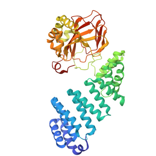
Structural basis of the promiscuity of the unusual Fe(II) and 2-oxoglutarate dependent human aspartate/asparagine-beta-hydroxylase.

de Munnik, M.Brasnett, A.Zhou, T.Myers, W.Wang, Y.Chatterjee, K.Tumber, A.Marshall, S.A.Simon, P.S.Aller, P.Shilova, A.Axford, D.Makita, H.Paley, D.W.Tiwari, V.Stead, A.T.Dehe, S.Sanchez, H.Rosenberg, D.J.Alonso-Mori, R.Bhowmick, A.Yano, J.Yachandra, V.K.Park, J.Park, S.Orville, A.M.Brewitz, L.Kern, J.F.Schofield, C.J.Rabe, P.

(2026) Nat Commun 

  • DOI: https://doi.org/10.1038/s41467-026-69425-w
  • Primary Citation of Related Structures:  
    9FVU, 9FVV, 9FVW, 9FVX, 9FVY, 9FVZ, 9FW0, 9HNZ, 9HO0, 9HO1, 9HO2, 9HO3

  • PubMed Abstract: 

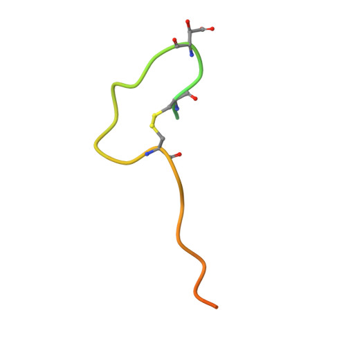
    Protein-hydroxylation catalysed by Fe(II) and 2-oxoglutarate (2OG) dependent oxygenases is an important regulatory mechanism in human biology. Such oxygenases typically coordinate their Fe(II) cofactor via a conserved triad of an aspartate- or glutamate- and two histidine-residues. By contrast, aspartate/asparagine β-hydroxylase (AspH), which catalyses asparagine/aspartate-residue oxidation in epidermal growth factor-like domains (EGFDs), has only two histidine-residues (H679, H725), with a water occupying the site normally occupied by an aspartate- or glutamate-residue. We describe mechanistic studies with catalytically active AspH crystals. Turnover studies with single crystals under cryogenic conditions give (3 R)-hydroxylated EGFDs with the product alcohol coordinating Fe(II) trans to H725. Time-resolved serial crystallography of microcrystals using an acoustic droplet ejection system, coupled to X-ray emission analyses, demonstrate turnover within 1.5 s, giving a product complex in which Fe(II) is regenerated. Solution and crystallographic studies with the O 2 surrogate nitric oxide imply O 2 binds to Fe(II) trans to H725. The additional Fe-chelating water is maintained throughout AspH catalysis and is not directly involved in substrate hydroxylation, because O 2 is the sole oxygen source in alcohol products, as shown by 18 O labelling studies. The results reveal how AspH accommodates both aspartate- and asparagine-substrates and will assist in efforts targeting AspH for cancer treatment.


  • Organizational Affiliation
    • Chemistry Research Laboratory and the Ineos Oxford Institute for Antimicrobial Research, University of Oxford, Oxford, UK.

Macromolecules
Find similar proteins by:  (by identity cutoff)  |  3D Structure
Entity ID: 1
MoleculeChains Sequence LengthOrganismDetailsImage
Aspartyl/asparaginyl beta-hydroxylase429Homo sapiensMutation(s): 0 
Gene Names: ASPHBAH
EC: 1.14.11.16
UniProt & NIH Common Fund Data Resources
Find proteins for Q12797 (Homo sapiens)
Explore Q12797 
Go to UniProtKB:  Q12797
PHAROS:  Q12797
GTEx:  ENSG00000198363 
Entity Groups  
Sequence Clusters30% Identity50% Identity70% Identity90% Identity95% Identity100% Identity
UniProt GroupQ12797
Sequence Annotations
Expand
  • Reference Sequence
Find similar proteins by:  (by identity cutoff)  |  3D Structure
Entity ID: 2
MoleculeChains Sequence LengthOrganismDetailsImage
Factor X light chain39Homo sapiensMutation(s): 4 
EC: 3.4.21.6
UniProt & NIH Common Fund Data Resources
Find proteins for P00742 (Homo sapiens)
Explore P00742 
Go to UniProtKB:  P00742
PHAROS:  P00742
GTEx:  ENSG00000126218 
Entity Groups  
Sequence Clusters30% Identity50% Identity70% Identity90% Identity95% Identity100% Identity
UniProt GroupP00742
Sequence Annotations
Expand
  • Reference Sequence
Small Molecules
Modified Residues  1 Unique
IDChains TypeFormula2D DiagramParent
BH2
Query on BH2
B
L-PEPTIDE LINKINGC4 H7 N O5ASP
Experimental Data & Validation

Experimental Data

  • Method: X-RAY DIFFRACTION
  • Resolution: 2.14 Å
  • R-Value Free:  0.232 (Depositor), 0.231 (DCC) 
  • R-Value Work:  0.208 (Depositor), 0.206 (DCC) 
  • R-Value Observed: 0.210 (Depositor) 
Space Group: P 21 21 21
Unit Cell:
Length ( Å )Angle ( ˚ )
a = 50.851α = 90
b = 91.441β = 90
c = 124.073γ = 90
Software Package:
Software NamePurpose
PHENIXrefinement
DIALSdata reduction
cctbx.xfel.mergedata scaling
PHENIXphasing

Structure Validation

View Full Validation Report


Entry History & Funding Information

Deposition Data


Funding OrganizationLocationGrant Number
Biotechnology and Biological Sciences Research Council (BBSRC)United KingdomBB/V003291/1

Revision History  (Full details and data files)

  • Version 1.0: 2025-12-24
    Type: Initial release
  • Version 1.1: 2026-03-11
    Changes: Database references