9HN5 | pdb_00009hn5

Cryo-EM structure of human separase bound to phosphorylated SCC1 (100-320 aa)


Experimental Data Snapshot

  • Method: ELECTRON MICROSCOPY
  • Resolution: 2.96 Å
  • Aggregation State: PARTICLE 
  • Reconstruction Method: SINGLE PARTICLE 

Starting Model: experimental
View more details

wwPDB Validation 3D Report Full Report

Validation slider image for 9HN5

This is version 1.1 of the entry. See complete history

Literature

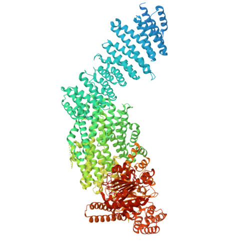
Substrate recognition by human separase.

Yu, J.Schmidt, S.Botto, M.Lee, K.Ghent, C.M.Goodfried, J.M.Howe, A.O'Reilly, F.J.Morgan, D.O.Boland, A.

(2025) Sci Adv 11: eady9807-eady9807

  • DOI: https://doi.org/10.1126/sciadv.ady9807
  • Primary Citation Related Structures: 
    9HM7, 9HMA, 9HMS, 9HMV, 9HN0, 9HN4, 9HN5

  • PubMed Abstract: 

    The cohesin complex encircles sister chromatids in early mitosis. At anaphase onset, sister separation is triggered by the proteolytic cleavage of the cohesin subunit SCC1/RAD21 by separase. SCC1 contains two cleavage sites, where cleavage is stimulated by SCC1 phosphorylation. Substrate recognition and cleavage are only partly understood. Here, we determined structures of human separase in apo- or substrate-bound forms that, together with biochemical analysis, provide critical insights into separase cleavage regulation. We verify the first SCC1 cleavage site and reassign the second. We show that substrates, including separase autocleavage sites and the two SCC1 cleavage sites, interact with docking sites in separase, including five phosphate-binding sites. We also describe the interaction between the cohesin subunit SA1/SA2 and separase, which promotes cleavage at the second SCC1 site. Using cross-linking mass spectrometry and cryo-electron microscopy, we propose how cohesin is targeted by human separase. Our work provides an extensive functional and structural framework that explains a key event in cell division.


  • Organizational Affiliation
    • Department of Molecular and Cellular Biology, University of Geneva, Geneva, Switzerland.

Macromolecule Content 

  • Total Structure Weight: 263.67 kDa 
  • Atom Count: 11,314 
  • Modeled Residue Count: 1,518 
  • Deposited Residue Count: 2,399 
  • Unique protein chains: 2

Macromolecules

Find similar proteins by:|  3D Structure
Entity ID: 1
MoleculeChains  Sequence LengthOrganismDetailsImage
Double-strand-break repair protein rad21 homologA [auth B]227Homo sapiensMutation(s): 0 
Gene Names: RAD21HR21KIAA0078NXP1SCC1
UniProt & NIH Common Fund Data Resources
Find proteins for O60216 (Homo sapiens)
Explore O60216 
Go to UniProtKB:  O60216
PHAROS:  O60216
GTEx:  ENSG00000164754 
Entity Groups
Sequence Clusters30% Identity50% Identity70% Identity90% Identity95% Identity100% Identity
UniProt GroupO60216
Sequence Annotations
Expand
Reference Sequence
Find similar proteins by:|  3D Structure
Entity ID: 2
MoleculeChains  Sequence LengthOrganismDetailsImage
SeparinB [auth A]2,172Homo sapiensMutation(s): 1 
Gene Names: ESPL1ESP1KIAA0165
EC: 3.4.22.49
UniProt & NIH Common Fund Data Resources
Find proteins for Q14674 (Homo sapiens)
Explore Q14674 
Go to UniProtKB:  Q14674
PHAROS:  Q14674
GTEx:  ENSG00000135476 
Entity Groups
Sequence Clusters30% Identity50% Identity70% Identity90% Identity95% Identity100% Identity
UniProt GroupQ14674
Sequence Annotations
Expand
Reference Sequence

Small Molecules

Modified Residues  1 Unique
IDChains TypeFormula2D DiagramParent
SEP
Query on SEP
A [auth B]L-PEPTIDE LINKINGC3 H8 N O6 PSER

Experimental Data & Validation

Experimental Data

  • Method: ELECTRON MICROSCOPY
  • Resolution: 2.96 Å
  • Aggregation State: PARTICLE 
  • Reconstruction Method: SINGLE PARTICLE 
EM Software:
TaskSoftware PackageVersion
MODEL REFINEMENTPHENIX1.20.1_4487
MODEL REFINEMENTCoot0.9.8.92
RECONSTRUCTIONcryoSPARC4.4.0

Structure Validation

View Full Validation Report


Entry History 

& Funding Information

Deposition Data


Funding OrganizationLocationGrant Number
Swiss National Science FoundationSwitzerland310030_185235
Swiss National Science FoundationSwitzerlandTMSGI3_211581

Revision History  (Full details and data files)

  • Version 1.0: 2025-09-03
    Type: Initial release
  • Version 1.1: 2026-05-06
    Changes: Data collection, Database references