9HMP | pdb_00009hmp

X-ray structure of S. cerevisiae threonylcarbamoyladenosine dehydratase 1 (residues 50-429) in complex with AMP


Experimental Data Snapshot

  • Method: X-RAY DIFFRACTION
  • Resolution: 4.00 Å
  • R-Value Free: 
    0.299 (Depositor), 0.290 (DCC) 
  • R-Value Work: 
    0.255 (Depositor), 0.249 (DCC) 
  • R-Value Observed: 
    0.257 (Depositor) 

Starting Model: in silico
View more details

wwPDB Validation 3D Report Full Report

Validation slider image for 9HMP

Ligand Structure Quality Assessment 


This is version 1.1 of the entry. See complete history

Literature

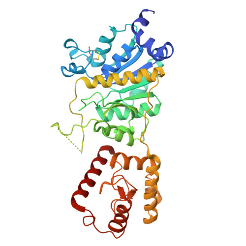
Structural and biochemical characterization of yeast Tcd enzymes installing the post-transcriptional modification ct6A in tRNA.

Hirschmann, J.Sonntag, R.Heiss, M.Wegrzyn, E.Heinemeyer, W.Carell, T.Huber, E.M.

(2026) Nucleic Acids Res 54

  • DOI: https://doi.org/10.1093/nar/gkag376
  • Primary Citation Related Structures: 
    9HMO, 9HMP, 9TZH

  • PubMed Abstract: 

    Post-transcriptional modifications near the anticodon of transfer ribonucleic acids (tRNAs) ensure translation fidelity and accuracy. For instance, at position 37, the universally conserved and essential nucleoside N6-threonylcarbamoyladenosine (t6A) supports decoding of ANN triplets. In some organisms t6A is converted to cyclic t6A (ct6A), but only little is known about this ATP-dependent reaction and the corresponding threonylcarbamoyladenosine dehydratases (Tcds). We here show that yeast Tcds localize to the outer mitochondrial membrane and co-purify with tRNAs recognizing ANN codons. Depending on the number of TCD genes in the genome, the proteins form V-shaped hetero- or homodimers, of which at least one subunit binds and modifies tRNAs. The C-terminal, monomeric domain shares similarities with Cas9-endonucleases and assists tRNA recognition, while the N-terminal domain mediates dimerization and contains the active site. Structure-based mutagenesis and activity assays imply that yeast Tcds lack a catalytic cysteine and do not covalently bind their substrate as proposed for Escherichia coli TcdA.


  • Organizational Affiliation
    • Technical University of Munich, TUM School of Natural Sciences, Center for Functional Protein Assemblies, 85747 Garching, Germany.

Macromolecule Content 

  • Total Structure Weight: 175.95 kDa 
  • Atom Count: 11,707 
  • Modeled Residue Count: 1,451 
  • Deposited Residue Count: 1,528 
  • Unique protein chains: 1

Macromolecules

Find similar proteins by:|  3D Structure
Entity ID: 1
MoleculeChains  Sequence LengthOrganismDetailsImage
tRNA threonylcarbamoyladenosine dehydratase 1
A, B, C, D
382Saccharomyces cerevisiaeMutation(s): 0 
Gene Names: TCD1YHR003C
EC: 6.1
UniProt
Find proteins for P38756 (Saccharomyces cerevisiae (strain ATCC 204508 / S288c))
Explore P38756 
Go to UniProtKB:  P38756
Entity Groups
Sequence Clusters30% Identity50% Identity70% Identity90% Identity95% Identity100% Identity
UniProt GroupP38756
Sequence Annotations
Expand
Reference Sequence

Small Molecules

Ligands 4 Unique
IDChains Name / Formula / InChI Key2D Diagram3D Interactions
AMP
(Subject of Investigation/LOI)

Query on AMP


Download:Ideal Coordinates CCD File
E [auth A],
K [auth B],
P [auth C],
T [auth D]
ADENOSINE MONOPHOSPHATE
C10 H14 N5 O7 P
UDMBCSSLTHHNCD-KQYNXXCUSA-N
GOL

Query on GOL


Download:Ideal Coordinates CCD File
L [auth B]GLYCEROL
C3 H8 O3
PEDCQBHIVMGVHV-UHFFFAOYSA-N
CL

Query on CL


Download:Ideal Coordinates CCD File
F [auth A],
G [auth A],
Q [auth C],
R [auth C],
U [auth D]
CHLORIDE ION
Cl
VEXZGXHMUGYJMC-UHFFFAOYSA-M
NA

Query on NA


Download:Ideal Coordinates CCD File
H [auth A]
I [auth A]
J [auth A]
M [auth B]
N [auth B]
H [auth A],
I [auth A],
J [auth A],
M [auth B],
N [auth B],
O [auth B],
S [auth C],
V [auth D]
SODIUM ION
Na
FKNQFGJONOIPTF-UHFFFAOYSA-N

Experimental Data & Validation

Experimental Data

  • Method: X-RAY DIFFRACTION
  • Resolution: 4.00 Å
  • R-Value Free:  0.299 (Depositor), 0.290 (DCC) 
  • R-Value Work:  0.255 (Depositor), 0.249 (DCC) 
  • R-Value Observed: 0.257 (Depositor) 
Space Group: P 65 2 2
Unit Cell:
Length ( Å )Angle ( ˚ )
a = 259.44α = 90
b = 259.44β = 90
c = 236.11γ = 120
Software Package:
Software NamePurpose
REFMACrefinement
XDSdata reduction
XSCALEdata scaling
PHASERphasing

Structure Validation

View Full Validation Report


Ligand Structure Quality Assessment 


Entry History 

& Funding Information

Deposition Data


Funding OrganizationLocationGrant Number
German Research Foundation (DFG)Germany325871075

Revision History  (Full details and data files)

  • Version 1.0: 2025-12-24
    Type: Initial release
  • Version 1.1: 2026-05-27
    Changes: Database references