


Crystal structure of PDE6D in complex with compound 5e
Zhang, R.To be published.
Experimental Data Snapshot
Starting Model: experimental
View more details
Entity ID: 1 | |||||
|---|---|---|---|---|---|
| Molecule | Chains | Sequence Length | Organism | Details | Image |
| Retinal rod rhodopsin-sensitive cGMP 3',5'-cyclic phosphodiesterase subunit delta | 156 | Homo sapiens | Mutation(s): 0 Gene Names: PDE6D, PDED | 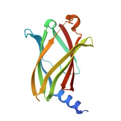 | |
UniProt & NIH Common Fund Data Resources | |||||
PHAROS: O43924 GTEx: ENSG00000156973 | |||||
Entity Groups | |||||
| Sequence Clusters | 30% Identity50% Identity70% Identity90% Identity95% Identity100% Identity | ||||
| UniProt Group | O43924 | ||||
Sequence AnnotationsExpand | |||||
Reference Sequence | |||||
| Ligands 3 Unique | |||||
|---|---|---|---|---|---|
| ID | Chains | Name / Formula / InChI Key | 2D Diagram | 3D Interactions | |
| A1IWC (Subject of Investigation/LOI) Download:Ideal Coordinates CCD File | C [auth A], F [auth B] | 4-[3,4-dimethyl-2-(4-methylphenyl)-7-oxidanylidene-pyrazolo[3,4-d]pyridazin-6-yl]-~{N}-heptyl-~{N}-(piperidin-4-ylmethyl)butane-1-sulfonamide C31 H48 N6 O3 S DVTXDCJEWQTSFM-UHFFFAOYSA-N |  | ||
| PEG Download:Ideal Coordinates CCD File | G [auth B] | DI(HYDROXYETHYL)ETHER C4 H10 O3 MTHSVFCYNBDYFN-UHFFFAOYSA-N |  | ||
| GOL Download:Ideal Coordinates CCD File | D [auth A], E [auth A] | GLYCEROL C3 H8 O3 PEDCQBHIVMGVHV-UHFFFAOYSA-N |  | ||
| Length ( Å ) | Angle ( ˚ ) |
|---|---|
| a = 56.74 | α = 90 |
| b = 75.65 | β = 90 |
| c = 82.31 | γ = 90 |
| Software Name | Purpose |
|---|---|
| PHENIX | refinement |
| XSCALE | data scaling |
| XDS | data reduction |
| PHASER | phasing |
| Funding Organization | Location | Grant Number |
|---|---|---|
| Not funded | -- |


