9HIZ | pdb_00009hiz

Complex of the Nanofitin Sac7d-C3(C24A) with a human IgG1 Fc fragment


Experimental Data Snapshot

  • Method: X-RAY DIFFRACTION
  • Resolution: 2.90 Å
  • R-Value Free: 
    0.279 (Depositor), 0.289 (DCC) 
  • R-Value Work: 
    0.227 (Depositor) 
  • R-Value Observed: 
    0.230 (Depositor) 

Starting Models: experimental
View more details

wwPDB Validation 3D Report Full Report

Validation slider image for 9HIZ

This is version 1.0 of the entry. See complete history

Literature

Engineering and Structural Elucidation of a Sac7d-Derived IgG Fc-Specific Affitin and Its Application for the Light-Controlled Affinity Purification of Antibodies.

Veitl, F.Eichinger, A.Mayrhofer, P.Anneser, M.R.Testanera, M.Rauscher, K.Lenz, M.Skerra, A.

(2025) Chembiochem 26: e202500102-e202500102

  • DOI: https://doi.org/10.1002/cbic.202500102
  • Primary Citation Related Structures: 
    9HIZ

  • PubMed Abstract: 

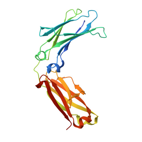
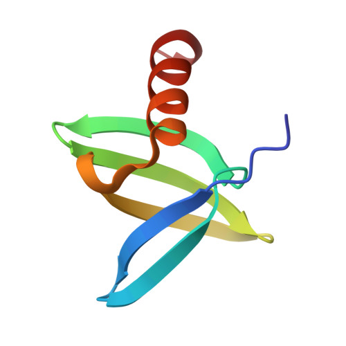
    While protein A affinity chromatography is widely established for antibody purification, the acidic elution conditions often lead to protein aggregation and deamidation. Here, an alternative approach is described for the purification of antibodies utilizing an engineered binding protein based on the archaebacterial Sac7d scaffold in combination with light-controlled α-CD affinity chromatography (Excitography). Starting from a published affitin molecule, a monomeric protein version (C3A24) was engineered by substituting the unpaired thiol side chain Cys24 within the binding site by Ala, and, unexpectedly, its binding activity towards the human IgG1 Fc region was even improved (K D  = 76 nM). X-ray analysis of the cocrystallized C3A24 with a recombinant human Fc fragment revealed a 2:1 stoichiometry, with a binding site at the junction between the C H 2 and C H 3 domains. Interestingly, this binding site coincides with the ones of protein A, protein G, and the neonatal Fc receptor (FcRn). The affitin/Fc interaction is dominated by a network of hydrogen bonds, whereas, unpredicted by the initial affitin design, the two C-terminal Lys residues are also involved via a salt bridge and another hydrogen bond. Using the Azo-tagged C3A24, we purified clinically relevant antibodies from cell culture medium in a single step under physiological buffer conditions.


  • Organizational Affiliation
    • Chair of Biological Chemistry, School of Life Sciences, Technical University of Munich, 85354, Freising, Germany.

Macromolecule Content 

  • Total Structure Weight: 130.68 kDa 
  • Atom Count: 8,892 
  • Modeled Residue Count: 1,112 
  • Deposited Residue Count: 1,144 
  • Unique protein chains: 2

Macromolecules

Find similar proteins by:|  3D Structure
Entity ID: 1
MoleculeChains  Sequence LengthOrganismDetailsImage
Immunoglobulin heavy constant gamma 1A,
B,
E [auth C],
F [auth D]
217Homo sapiensMutation(s): 0 
Gene Names: IGHG1
UniProt & NIH Common Fund Data Resources
Find proteins for P01857 (Homo sapiens)
Explore P01857 
Go to UniProtKB:  P01857
PHAROS:  P01857
GTEx:  ENSG00000211896 
Entity Groups
Sequence Clusters30% Identity50% Identity70% Identity90% Identity95% Identity100% Identity
UniProt GroupP01857
Sequence Annotations
Expand
Reference Sequence
Find similar proteins by:|  3D Structure
Entity ID: 2
MoleculeChains  Sequence LengthOrganismDetailsImage
DNA-binding protein 7dC [auth R],
D [auth S],
G [auth T],
H [auth U]
69Sulfolobus acidocaldarius DSM 639Mutation(s): 15 
Gene Names: Saci_0064
UniProt
Find proteins for P13123 (Sulfolobus acidocaldarius (strain ATCC 33909 / DSM 639 / JCM 8929 / NBRC 15157 / NCIMB 11770))
Explore P13123 
Go to UniProtKB:  P13123
Entity Groups
Sequence Clusters30% Identity50% Identity70% Identity90% Identity95% Identity100% Identity
UniProt GroupP13123
Sequence Annotations
Expand
Reference Sequence

Experimental Data & Validation

Experimental Data

  • Method: X-RAY DIFFRACTION
  • Resolution: 2.90 Å
  • R-Value Free:  0.279 (Depositor), 0.289 (DCC) 
  • R-Value Work:  0.227 (Depositor) 
  • R-Value Observed: 0.230 (Depositor) 
Space Group: P 1 21 1
Unit Cell:
Length ( Å )Angle ( ˚ )
a = 68.27α = 90
b = 136.84β = 90.12
c = 69.76γ = 90
Software Package:
Software NamePurpose
REFMACrefinement
XDSdata scaling
XDSdata reduction
PHASERphasing

Structure Validation

View Full Validation Report


Entry History 

& Funding Information

Deposition Data


Funding OrganizationLocationGrant Number
Not funded--

Revision History  (Full details and data files)

  • Version 1.0: 2025-07-23
    Type: Initial release