9HIX | pdb_00009hix

Cryo-EM structure of CAK (CDK7 D97N mutant) in complex with THZ1


Experimental Data Snapshot

  • Method: ELECTRON MICROSCOPY
  • Resolution: 2.60 Å
  • Aggregation State: PARTICLE 
  • Reconstruction Method: SINGLE PARTICLE 

Starting Model: experimental
View more details

wwPDB Validation 3D Report Full Report

Validation slider image for 9HIX

This is version 1.1 of the entry. See complete history

Literature

Resistance to CDK7 inhibitors directed by acquired mutation of a conserved residue in cancer cells.

Lai, C.F.Cushing, V.I.Olden, E.Bevan, C.L.Coombes, R.C.Greber, B.J.Buluwela, L.Ali, S.

(2025) EMBO J 44: 5860-5889

  • DOI: https://doi.org/10.1038/s44318-025-00554-6
  • Primary Citation Related Structures: 
    9HIX, 9HIY, 9HJ0

  • PubMed Abstract: 

    CDK7 has emerged as a cancer target because of its pivotal roles in cell cycle progression and transcription. Several CDK7 inhibitors (CDK7i) are now in clinical evaluation. Identifying patients most likely to respond to treatment and early detection of tumour evolution towards resistance are necessary for optimal implementation of cancer therapies. Continuous culturing of prostate cancer cells with Samuraciclib, a non-covalent ATP-competitive CDK7i, led to outgrowth of resistant cells. These were characterised by the acquisition of a single base change in the CDK7 gene, Asp97 to Asn (D97N). Mutant cells were resistant to other non-covalent CDK7i but remained sensitive to covalent CDK7i. Cryo-EM structure and kinase ligand affinity determinations revealed reduced affinity of the CDK7-D97N mutant for non-covalent CDK7i. Remarkably, Asp97 is absolutely conserved in human CDKs, inferring its importance for the activities of all CDKs. Consistent with this, mutation of the homologous residue in CDK12 (D819N) or CDK4 (D99N) promoted resistance to drugs that inhibit these CDKs. Our findings reveal a general mechanism for acquired resistance with obvious implications for patients treated with CDK inhibitors.


  • Organizational Affiliation
    • Department of Surgery & Cancer, Imperial, London, UK.

Macromolecule Content 

  • Total Structure Weight: 87.89 kDa 
  • Atom Count: 5,180 
  • Modeled Residue Count: 626 
  • Deposited Residue Count: 766 
  • Unique protein chains: 3

Macromolecules

Find similar proteins by:|  3D Structure
Entity ID: 1
MoleculeChains  Sequence LengthOrganismDetailsImage
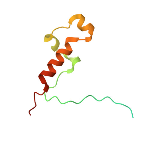
CDK-activating kinase assembly factor MAT1A [auth H]93Homo sapiensMutation(s): 0 
Gene Names: MNAT1CAP35MAT1RNF66
UniProt & NIH Common Fund Data Resources
Find proteins for P51948 (Homo sapiens)
Explore P51948 
Go to UniProtKB:  P51948
PHAROS:  P51948
GTEx:  ENSG00000020426 
Entity Groups
Sequence Clusters30% Identity50% Identity70% Identity90% Identity95% Identity100% Identity
UniProt GroupP51948
Sequence Annotations
Expand
Reference Sequence
Find similar proteins by:|  3D Structure
Entity ID: 2
MoleculeChains  Sequence LengthOrganismDetailsImage
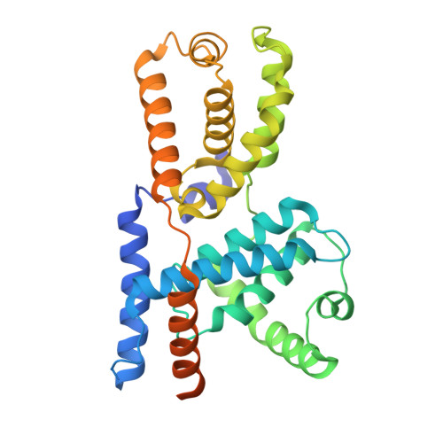
Cyclin-HB [auth I]324Homo sapiensMutation(s): 1 
Gene Names: CCNH
UniProt & NIH Common Fund Data Resources
Find proteins for P51946 (Homo sapiens)
Explore P51946 
Go to UniProtKB:  P51946
PHAROS:  P51946
GTEx:  ENSG00000134480 
Entity Groups
Sequence Clusters30% Identity50% Identity70% Identity90% Identity95% Identity100% Identity
UniProt GroupP51946
Sequence Annotations
Expand
Reference Sequence
Find similar proteins by:|  3D Structure
Entity ID: 3
MoleculeChains  Sequence LengthOrganismDetailsImage
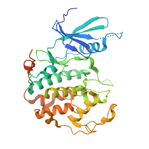
Cyclin-dependent kinase 7C [auth J]349Homo sapiensMutation(s): 1 
Gene Names: CDK7CAKCAK1CDKN7MO15STK1
EC: 2.7.11.22 (PDB Primary Data), 2.7.11.23 (PDB Primary Data)
UniProt & NIH Common Fund Data Resources
Find proteins for P50613 (Homo sapiens)
Explore P50613 
Go to UniProtKB:  P50613
PHAROS:  P50613
GTEx:  ENSG00000134058 
Entity Groups
Sequence Clusters30% Identity50% Identity70% Identity90% Identity95% Identity100% Identity
UniProt GroupP50613
Sequence Annotations
Expand
Reference Sequence

Small Molecules

Ligands 1 Unique
IDChains Name / Formula / InChI Key2D Diagram3D Interactions
V0G
(Subject of Investigation/LOI)

Query on V0G


Download:Ideal Coordinates CCD File
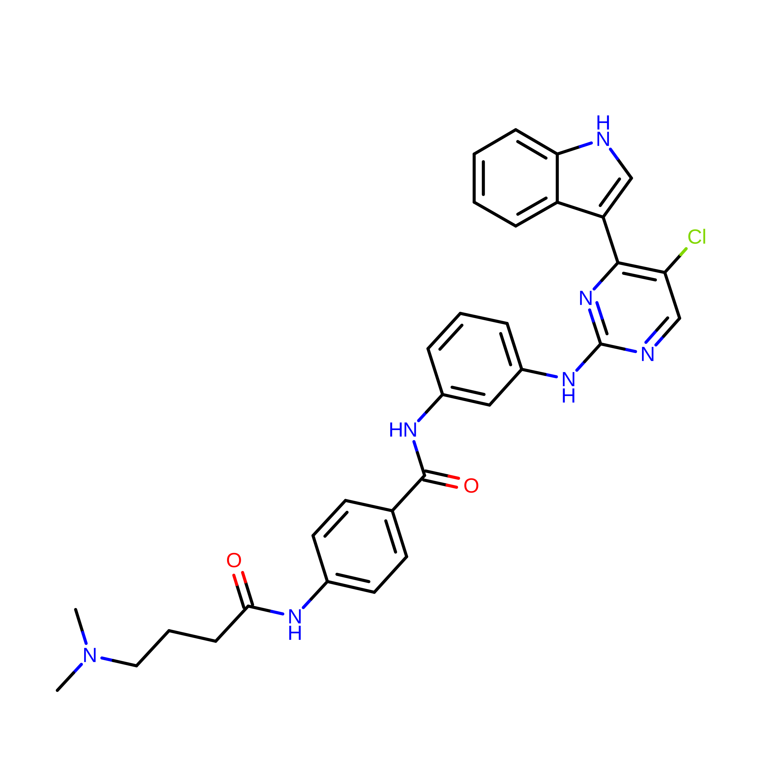
D [auth J]N-(3-{[5-chloro-4-(1H-indol-3-yl)pyrimidin-2-yl]amino}phenyl)-4-{[4-(dimethylamino)butanoyl]amino}benzamide
C31 H30 Cl N7 O2
TUERFPPIPKZNKE-UHFFFAOYSA-N

Experimental Data & Validation

Experimental Data

  • Method: ELECTRON MICROSCOPY
  • Resolution: 2.60 Å
  • Aggregation State: PARTICLE 
  • Reconstruction Method: SINGLE PARTICLE 
EM Software:
TaskSoftware PackageVersion
RECONSTRUCTIONRELION5.0
MODEL REFINEMENTPHENIX1.21_5207

Structure Validation

View Full Validation Report


Entry History 

& Funding Information

Deposition Data


Funding OrganizationLocationGrant Number
Medical Research Council (MRC, United Kingdom)United KingdomMR/V009354/1

Revision History  (Full details and data files)

  • Version 1.0: 2025-08-20
    Type: Initial release
  • Version 1.1: 2026-09-02
    Changes: Data collection, Database references