Deciphering the RNA recognition by Musashi-1 to design protein and RNA variants for in vitro and in vivo applications.
Perez-Rafols, A., Perez-Ropero, G., Cerofolini, L., Sperotto, L., Roca-Martinez, J., Higuera-Rodriguez, R.A., Russomanno, P., Kaiser, W., Vranken, W., Danielson, U.H., Provenzani, A., Martelli, T., Sattler, M., Buijs, J., Fragai, M.(2025) Nucleic Acids Res 53
- PubMed: 40795964 Search on PubMedSearch on PubMed Central
- DOI: https://doi.org/10.1093/nar/gkaf741
- Primary Citation Related Structures:
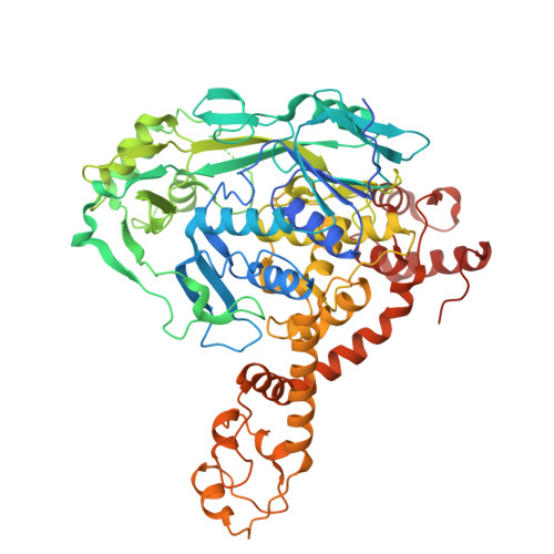
9HIR - PubMed Abstract:
The Human Musashi-1 (MSI-1) is an RNA-binding protein that recognizes (G/A)U1-3AGU and UAG sequences in diverse RNAs through two RNA Recognition Motif (RRM) domains and regulates the fate of target RNA. Here, we have combined structural biology and computational approaches to analyse the binding of the RRM domains of human MSI-1 with single-stranded and structured RNA ligands. We have used our recently developed computational tool RRMScorer to design a set of substitutions in the MSI-1 protein and the investigated RNA strands to modulate the binding affinity and selectivity. The in silico predictions of the designed protein-RNA interactions are assessed by nuclear magnetic resonance and surface plasmon resonance. These experiments have also been used to study the competition of the two RRM domains of MSI-1 for the same binding site within linear and harpin RNA. Our experimental results shed light on MSI-RNA interactions, thus opening the way for the development of new biomolecules for in vitro and in vivo studies and downstream applications.
- Magnetic Resonance Center (CERM) and Department of Chemistry, University of Florence, and Consorzio Interuniversitario Risonanze Magnetiche di Metalloproteine (CIRMMP), Sesto Fiorentino 50019, FI, Italy.
Organizational Affiliation: