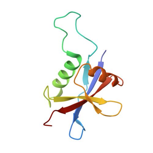
Structural organization of p62 filaments and the cellular ultrastructure of calcium-rich p62-enwrapped lipid droplet cargo.
Berkamp, S., Jungbluth, L., Katranidis, A., Mostafavi, S., Korculanin, O., Lu, P.H., Ickert, L., Dierig, M.M., Sharma, L., Thukral, L., Huesgen, P.F., Kononenko, N.L., Fitter, J., Dunin-Borkowski, R.E., Sachse, C.(2025) Nat Commun 16: 10810-10810
- PubMed: 41315362 Search on PubMedSearch on PubMed Central
- DOI: https://doi.org/10.1038/s41467-025-66785-7
- Primary Citation Related Structures:
9HGE - PubMed Abstract:
The selective autophagy receptor p62/SQSTM1 is known to form higher-order filaments in vitro and to undergo liquid-liquid phase separation when mixed with poly-ubiquitin. Here, we determine the full-length cryo-EM structure of p62 and elucidate a structured double helical filament scaffold composed of the PB1-domain associated with the flexible C-terminal part and the solvent-accessible major groove. At different pH values and upon binding to soluble LC3, LC3-conjugated membranes and poly-ubiquitin, we observe p62 filament re-arrangements in the form of structural unwinding, disassembly, lateral association and bundling, respectively. In the cellular environment, under conditions of ATG5 knockdown leading to stalled autophagy, we imaged high-contrast layers consisting of p62 oligomers enwrapping lipid droplets by cryogenic electron tomography in situ, which we identified as calcium as well as phosphorus by compositional spectroscopy analysis. Together, we visualize the cellular ultrastructure of p62 oligomers with high calcium content as a potential early stage of autophagy.
- Ernst-Ruska Centre for Microscopy and Spectroscopy with Electrons, ER-C-3/Structural Biology, Forschungszentrum Jülich, Jülich, Germany. s.berkamp@fz-juelich.de.
Organizational Affiliation: