9HDF | pdb_00009hdf

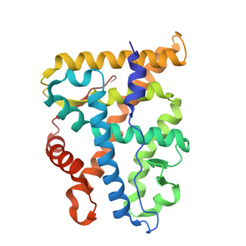
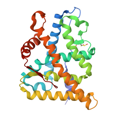
Glucocorticoid Receptor Ligand Binding Domain in complex with dexamethasone


Experimental Data Snapshot

  • Method: X-RAY DIFFRACTION
  • Resolution: 2.78 Å
  • R-Value Free: 
    0.244 (Depositor), 0.245 (DCC) 
  • R-Value Work: 
    0.211 (Depositor), 0.211 (DCC) 
  • R-Value Observed: 
    0.212 (Depositor) 

Starting Model: experimental
View more details

wwPDB Validation 3D Report Full Report

Validation slider image for 9HDF

Ligand Structure Quality Assessment 


This is version 1.0 of the entry. See complete history

Literature

The multimerization pathway of the glucocorticoid receptor.

Alegre-Marti, A.Jimenez-Panizo, A.Lafuente, A.L.Johnson, T.A.Montoya-Novoa, I.Peralta-Moreno, M.N.Montanya-Valluguera, P.Ponseti-Pons, J.Abella, M.Kim, S.Diaz, M.Vilaseca, M.Perez, P.Fernandez-Recio, J.Rubio-Martinez, J.Presman, D.M.Hager, G.L.Fuentes-Prior, P.Estebanez-Perpina, E.

(2025) Nucleic Acids Res 53

  • DOI: https://doi.org/10.1093/nar/gkaf1003
  • Primary Citation Related Structures: 
    9HDF

  • PubMed Abstract: 

    The glucocorticoid receptor (GR) is a leading drug target due to its antiinflammatory and immunosuppressive roles. The functional oligomeric conformation of full-length GR (FL-GR), which is key for its biological activity, remains disputed. Here we present a new crystal structure of agonist-bound GR ligand-binding domain (GR-LBD) comprising eight copies of a noncanonical dimer. We verified the biological relevance of this dimer for receptor multimerization in wild-type and selected FL-GR mutants using molecular dynamics and crosslinking-mass spectrometry together with fluorescence microscopy and transcriptomic analysis in living cells. Self-association of this GR-LBD basic dimer in two mutually exclusive assemblies reveals clues for FL-GR multimerization and activity in cells. We propose a model for the structure of multidomain GR based on our new data and suggest a detailed oligomerization pathway. This model reconciles all currently available structural and functional information and provides a more comprehensive understanding of the rare disorder, generalized glucocorticoid resistance.


  • Organizational Affiliation
    • Department of Biochemistry and Molecular Biomedicine, Faculty of Biology, University of Barcelona (UB), Barcelona 08028, Spain.

Macromolecule Content 

  • Total Structure Weight: 497.63 kDa 
  • Atom Count: 34,585 
  • Modeled Residue Count: 4,169 
  • Deposited Residue Count: 4,208 
  • Unique protein chains: 3

Macromolecules

Find similar proteins by:|  3D Structure
Entity ID: 1
MoleculeChains  Sequence LengthOrganismDetailsImage
Ancestral Glucocorticoid Receptor2 ligand binding domain
A, C, D, F, G
A, C, D, F, G, H, I, L, M, P
248Homo sapiensMutation(s): 0 
Find similar proteins by:|  3D Structure
Entity ID: 2
MoleculeChains  Sequence LengthOrganismDetailsImage
Ancestral Glucocorticoid Receptor2 ligand binding domain
B, E, J, K, N
B, E, J, K, N, O
248Homo sapiensMutation(s): 0 
Find similar proteins by:  Sequence   |   3D Structure  
Entity ID: 3
MoleculeChains  Sequence LengthOrganismDetailsImage
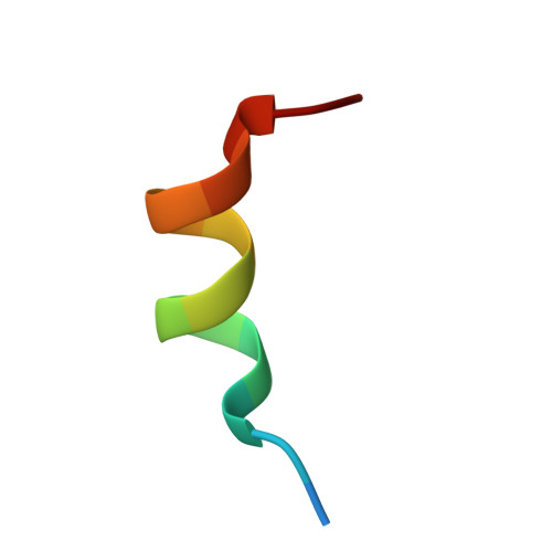
Nuclear receptor subfamily 0 group B member 215Homo sapiensMutation(s): 0 
UniProt & NIH Common Fund Data Resources
Find proteins for Q15466 (Homo sapiens)
Explore Q15466 
Go to UniProtKB:  Q15466
PHAROS:  Q15466
GTEx:  ENSG00000131910 
Entity Groups
Sequence Clusters30% Identity50% Identity70% Identity90% Identity95% Identity100% Identity
UniProt GroupQ15466
Sequence Annotations
Expand
Reference Sequence

Small Molecules

Ligands 10 Unique
IDChains Name / Formula / InChI Key2D Diagram3D Interactions
DEX

Query on DEX


Download:Ideal Coordinates CCD File
AD [auth M]
EB [auth D]
EC [auth J]
GA [auth A]
GD [auth N]
AD [auth M],
EB [auth D],
EC [auth J],
GA [auth A],
GD [auth N],
IB [auth E],
KD [auth O],
NA [auth B],
NC [auth K],
OD [auth P],
PB [auth F],
RC [auth L],
TB [auth G],
VB [auth H],
WA [auth C],
XB [auth I]
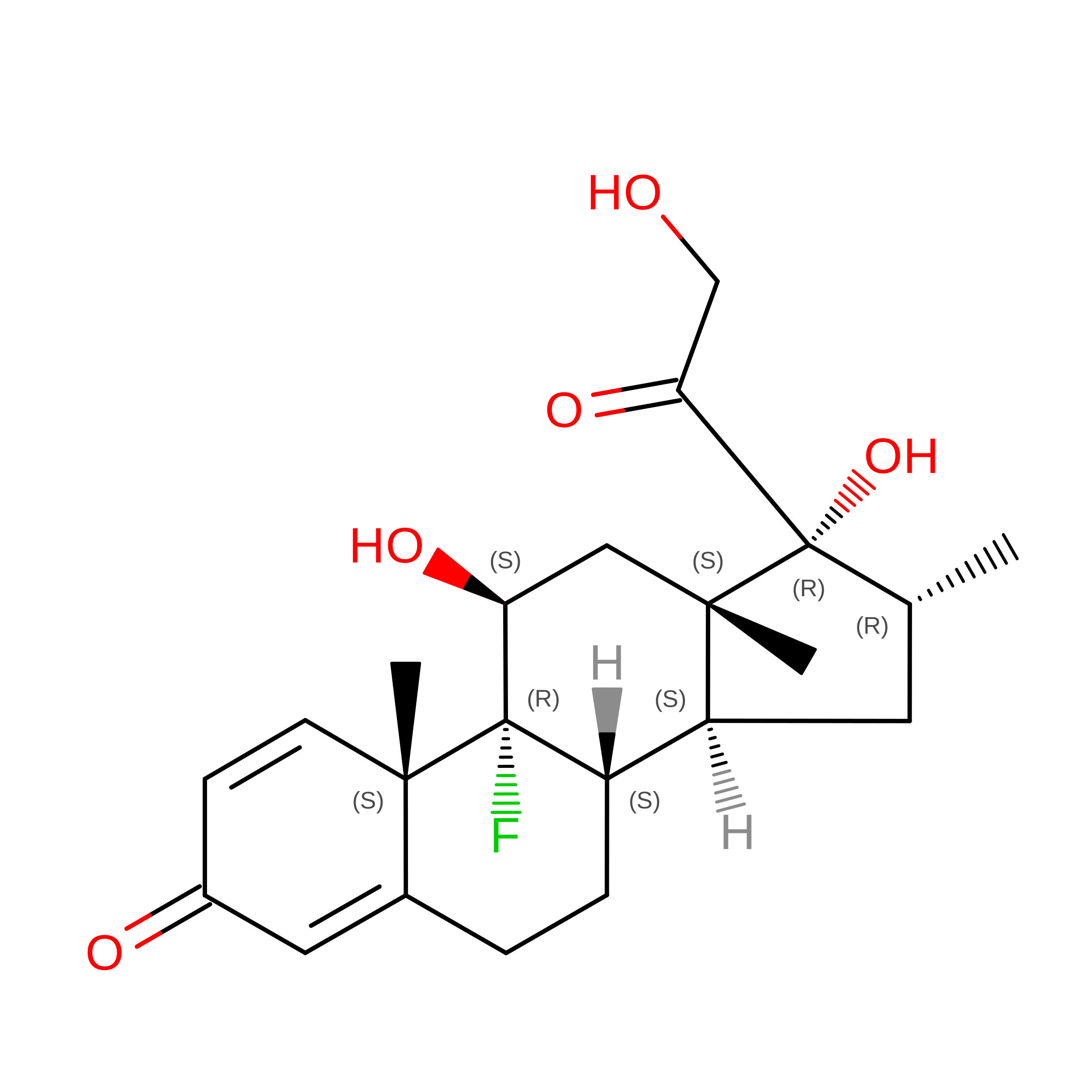
DEXAMETHASONE
C22 H29 F O5
UREBDLICKHMUKA-CXSFZGCWSA-N
EPE

Query on EPE


Download:Ideal Coordinates CCD File
AC [auth I]
FC [auth J]
GB [auth D]
IA [auth A]
PA [auth B]
AC [auth I],
FC [auth J],
GB [auth D],
IA [auth A],
PA [auth B],
PD [auth P],
TC [auth L]
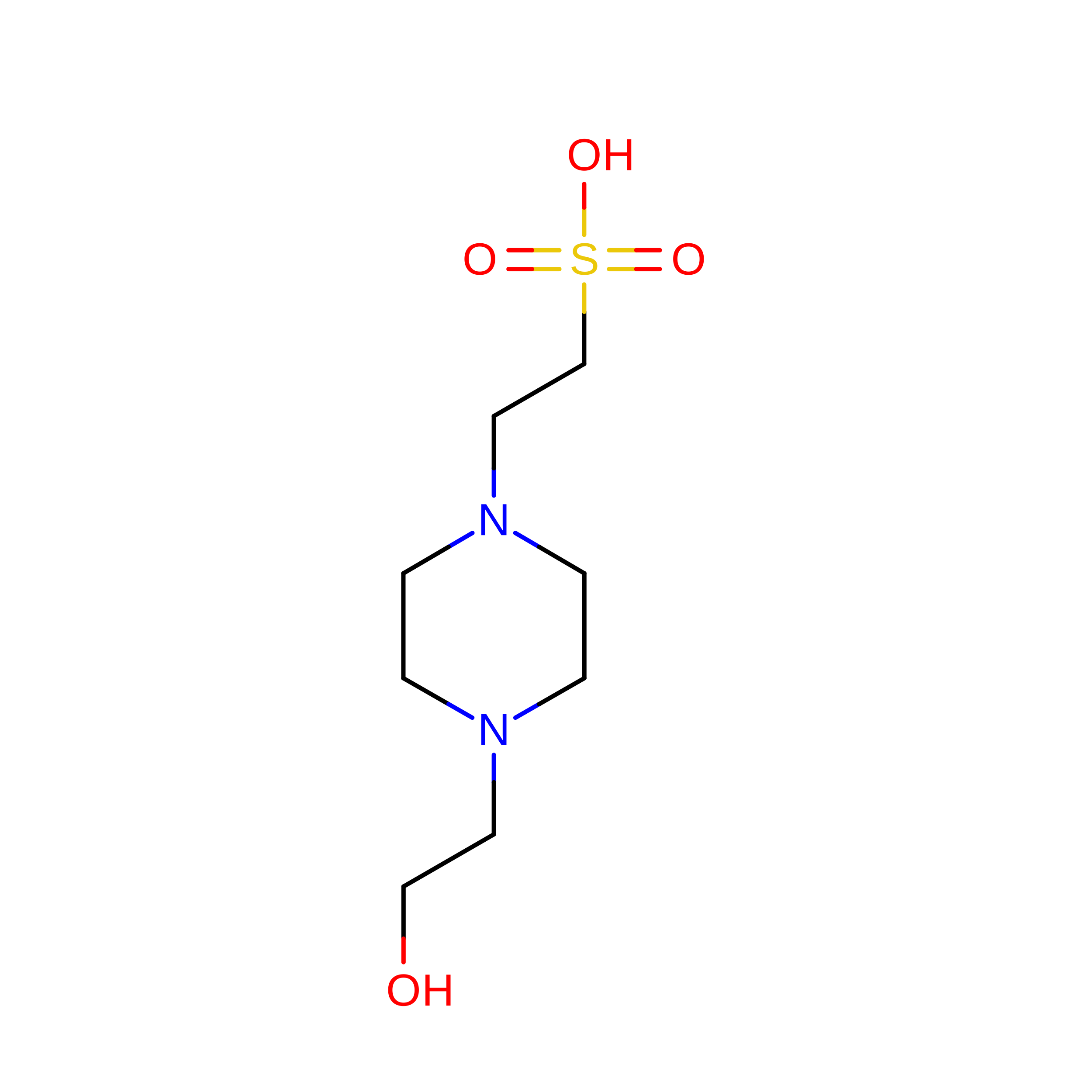
4-(2-HYDROXYETHYL)-1-PIPERAZINE ETHANESULFONIC ACID
C8 H18 N2 O4 S
JKMHFZQWWAIEOD-UHFFFAOYSA-N
PG4

Query on PG4


Download:Ideal Coordinates CCD File
FB [auth D],
OA [auth B],
SC [auth L],
WB [auth H]
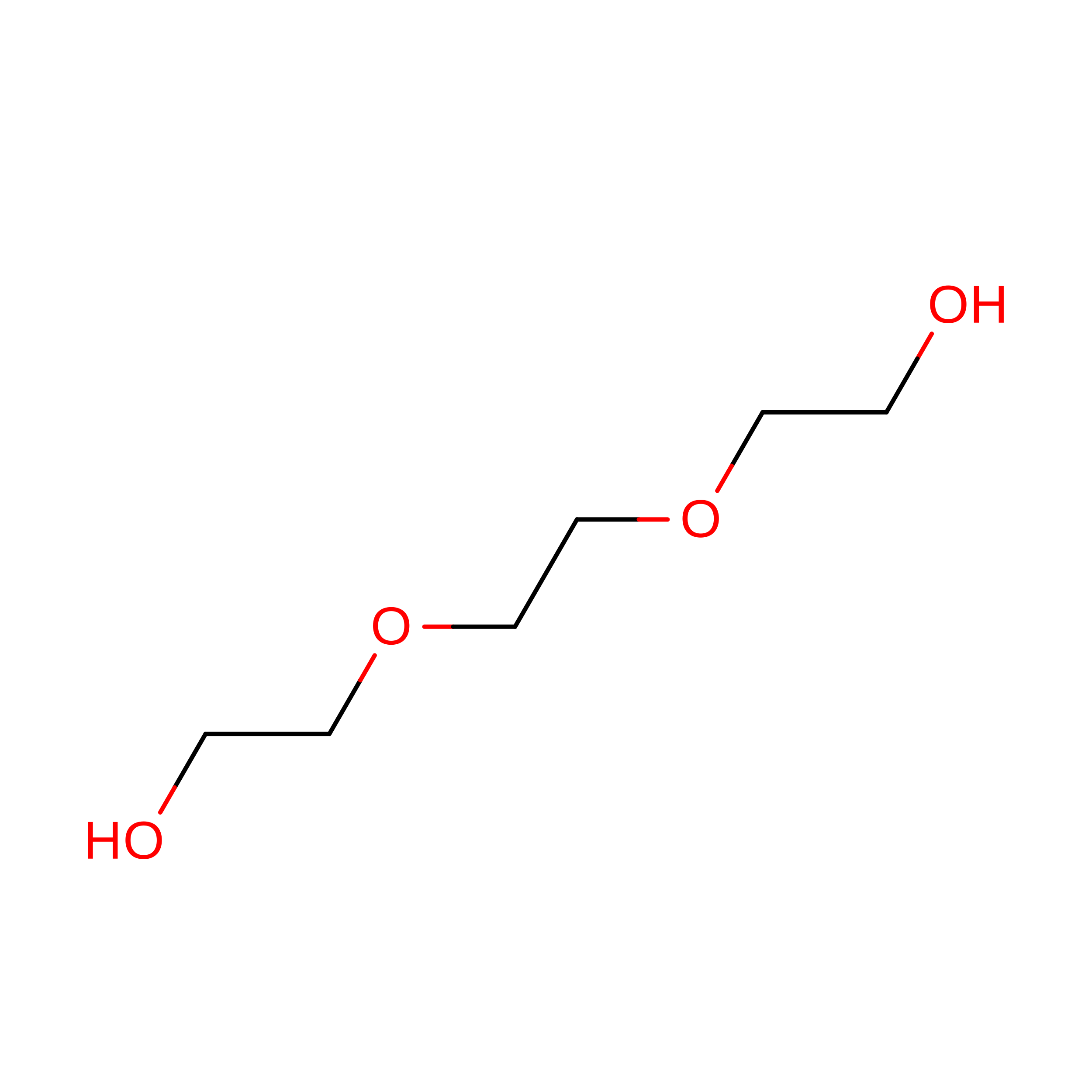
TETRAETHYLENE GLYCOL
C8 H18 O5
UWHCKJMYHZGTIT-UHFFFAOYSA-N
PGE

Query on PGE


Download:Ideal Coordinates CCD File
CD [auth M]TRIETHYLENE GLYCOL
C6 H14 O4
ZIBGPFATKBEMQZ-UHFFFAOYSA-N
CAC

Query on CAC


Download:Ideal Coordinates CCD File
BD [auth M]
DB [auth D]
HA [auth A]
JB [auth E]
LD [auth O]
BD [auth M],
DB [auth D],
HA [auth A],
JB [auth E],
LD [auth O],
OC [auth K],
UB [auth H],
YB [auth I]
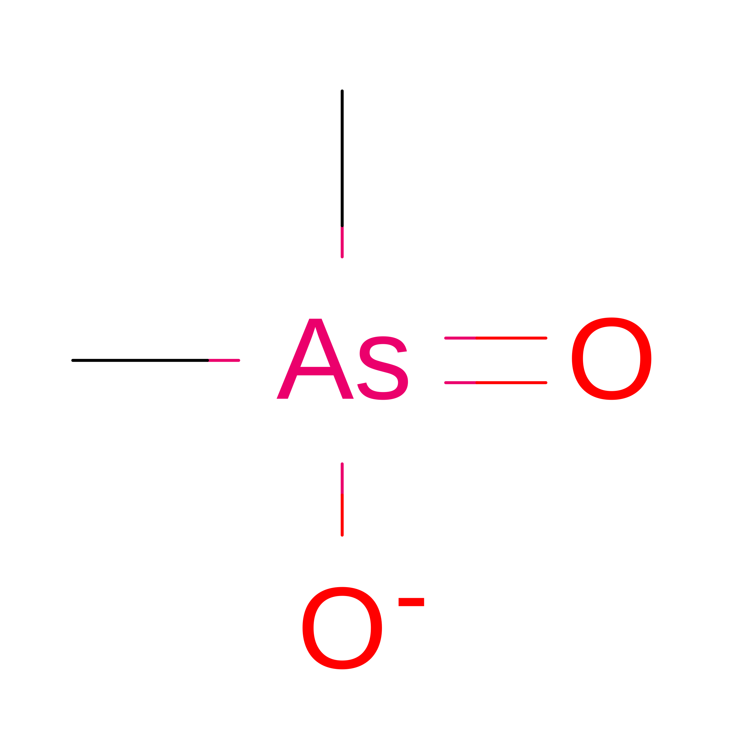
CACODYLATE ION
C2 H6 As O2
OGGXGZAMXPVRFZ-UHFFFAOYSA-M
PEG

Query on PEG


Download:Ideal Coordinates CCD File
DD [auth M],
MC [auth K],
QB [auth F],
ZB [auth I]
DI(HYDROXYETHYL)ETHER
C4 H10 O3
MTHSVFCYNBDYFN-UHFFFAOYSA-N
SO4

Query on SO4


Download:Ideal Coordinates CCD File
AB [auth C],
BB [auth C],
DC [auth J],
LA [auth A],
TA [auth B]
SULFATE ION
O4 S
QAOWNCQODCNURD-UHFFFAOYSA-L
GOL

Query on GOL


Download:Ideal Coordinates CCD File
BC [auth I]
GC [auth J]
HC [auth J]
HD [auth N]
IC [auth J]
BC [auth I],
GC [auth J],
HC [auth J],
HD [auth N],
IC [auth J],
JA [auth A],
KA [auth A],
KB [auth E],
LB [auth E],
MB [auth E],
MD [auth O],
PC [auth K],
QA [auth B],
QC [auth K],
QD [auth P],
UC [auth L],
VC [auth L],
XA [auth C],
YA [auth C],
ZC [auth M]
GLYCEROL
C3 H8 O3
PEDCQBHIVMGVHV-UHFFFAOYSA-N
IMD

Query on IMD


Download:Ideal Coordinates CCD File
CC [auth I]
ED [auth M]
ID [auth N]
JC [auth J]
KC [auth J]
CC [auth I],
ED [auth M],
ID [auth N],
JC [auth J],
KC [auth J],
NB [auth E],
ND [auth O],
RA [auth B],
RB [auth F],
RD [auth b],
SA [auth B],
SD [auth d],
WC [auth L],
XC [auth L],
ZA [auth C]
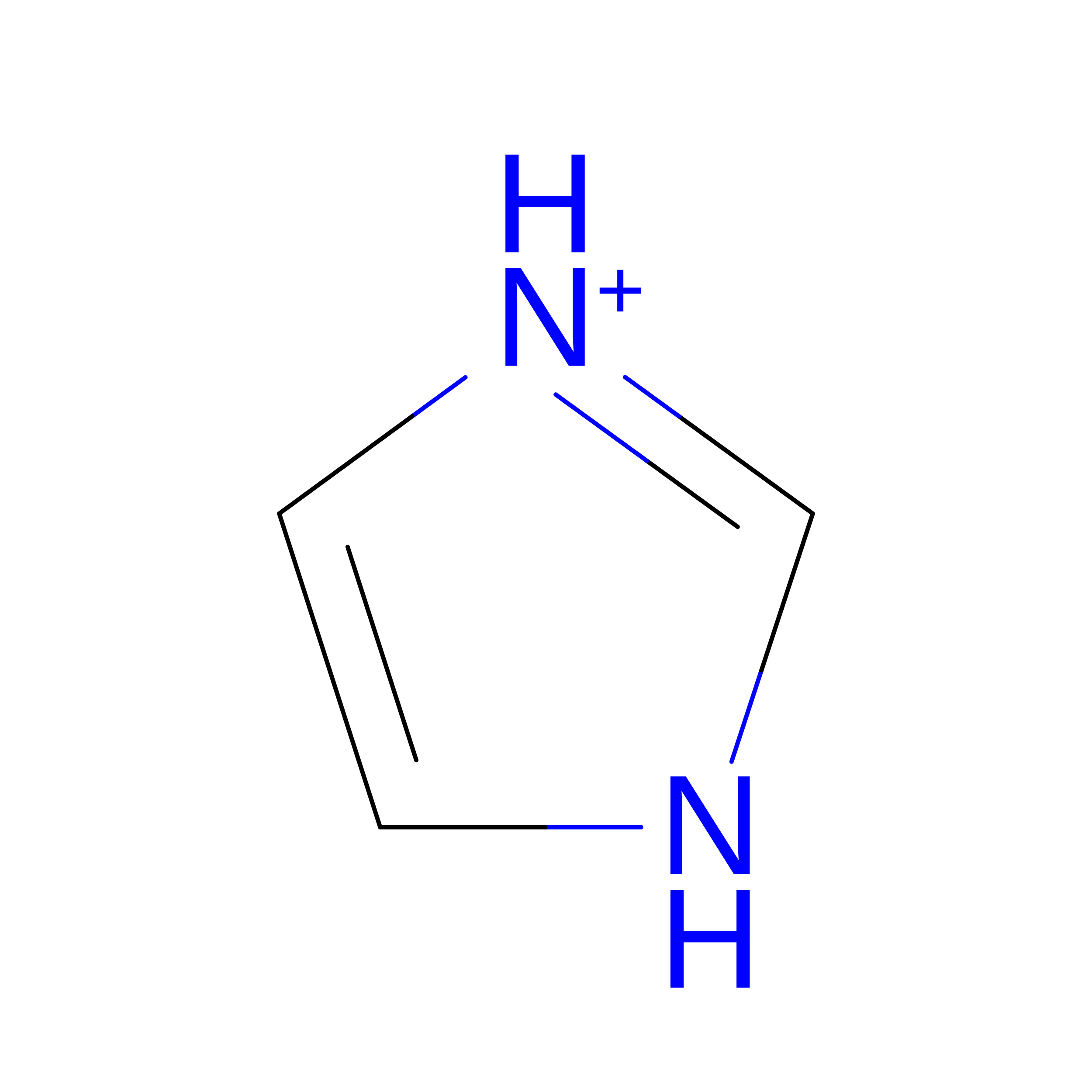
IMIDAZOLE
C3 H5 N2
RAXXELZNTBOGNW-UHFFFAOYSA-O
CL

Query on CL


Download:Ideal Coordinates CCD File
CB [auth C]
FD [auth M]
HB [auth D]
JD [auth N]
LC [auth J]
CB [auth C],
FD [auth M],
HB [auth D],
JD [auth N],
LC [auth J],
MA [auth B],
OB [auth E],
SB [auth F],
UA [auth B],
VA [auth B],
YC [auth L]
CHLORIDE ION
Cl
VEXZGXHMUGYJMC-UHFFFAOYSA-M
Modified Residues  1 Unique
IDChains TypeFormula2D DiagramParent
CSO
Query on CSO
B, E, J, K, N
B, E, J, K, N, O
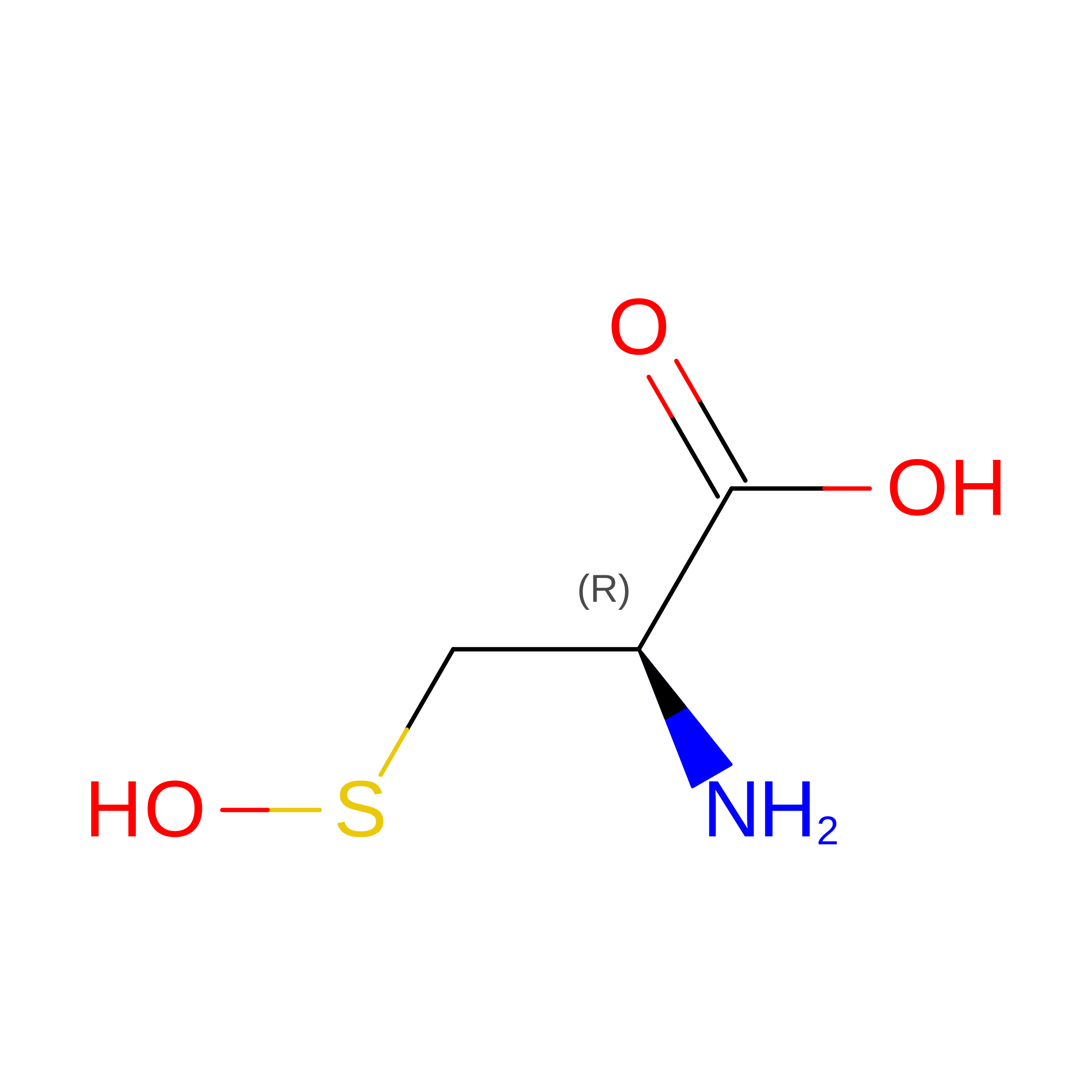
L-PEPTIDE LINKINGC3 H7 N O3 SCYS

Experimental Data & Validation

Experimental Data

  • Method: X-RAY DIFFRACTION
  • Resolution: 2.78 Å
  • R-Value Free:  0.244 (Depositor), 0.245 (DCC) 
  • R-Value Work:  0.211 (Depositor), 0.211 (DCC) 
  • R-Value Observed: 0.212 (Depositor) 
Space Group: P 21 21 2
Unit Cell:
Length ( Å )Angle ( ˚ )
a = 264.41α = 90
b = 265.464β = 90
c = 109.671γ = 90
Software Package:
Software NamePurpose
REFMACrefinement
Aimlessdata scaling
iMOSFLMdata reduction
MOLREPphasing

Structure Validation

View Full Validation Report


Ligand Structure Quality Assessment 


Entry History 

& Funding Information

Deposition Data


Funding OrganizationLocationGrant Number
Not funded--

Revision History  (Full details and data files)

  • Version 1.0: 2025-11-12
    Type: Initial release