


Crystal structure of a chimeric PPM1H phosphatase with the flap domain of PPM1J
Khan, A.R.To be published.
Experimental Data Snapshot
Starting Model: experimental
View more details
wwPDB Validation 3D Report Full Report

Entity ID: 1 | |||||
|---|---|---|---|---|---|
| Molecule | Chains | Sequence Length | Organism | Details | Image |
| PPM1H metal-dependent Ser/Thr phosphatase | 451 | Homo sapiens | Mutation(s): 0 EC: 3.1.3.16 | 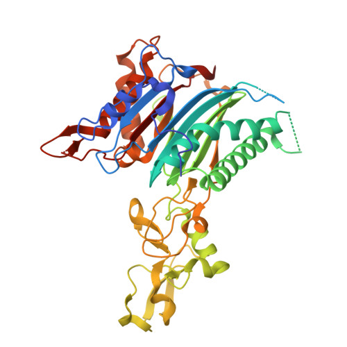 | |
| Ligands 1 Unique | |||||
|---|---|---|---|---|---|
| ID | Chains | Name / Formula / InChI Key | 2D Diagram | 3D Interactions | |
| MN (Subject of Investigation/LOI) Download:Ideal Coordinates CCD File | B [auth A], C [auth A], D [auth A] | MANGANESE (II) ION Mn WAEMQWOKJMHJLA-UHFFFAOYSA-N |  | ||
| Length ( Å ) | Angle ( ˚ ) |
|---|---|
| a = 110.093 | α = 90 |
| b = 110.093 | β = 90 |
| c = 87.597 | γ = 90 |
| Software Name | Purpose |
|---|---|
| PHENIX | refinement |
| Aimless | data scaling |
| XDS | data reduction |
| PHASER | phasing |
| Funding Organization | Location | Grant Number |
|---|---|---|
| Science Foundation Ireland | Ireland | 20/FFP-A/8446 |


