Twelve phosphomimetic mutations induce the assembly of recombinant full-length human tau into paired helical filaments.
Lovestam, S., Wagstaff, J.L., Katsinelos, T., Shi, J., Freund, S.M.V., Goedert, M., Scheres, S.H.W.(2026) Elife 14
- PubMed: 42159330 Search on PubMedSearch on PubMed Central
- DOI: https://doi.org/10.7554/eLife.104778
- Primary Citation Related Structures:
9H5G, 9H5J, 9S2B - PubMed Abstract:
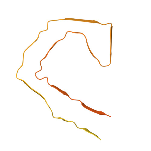
The assembly of tau into amyloid filaments is associated with more than 20 neurodegenerative diseases, collectively termed tauopathies. Electron cryo-microscopy (cryo-EM) structures of brain-derived tau filaments revealed that specific structures define different diseases, triggering a quest for the development of experimental model systems that replicate the structures of disease. Here, we describe 12 phosphomimetic serine/threonine-to-aspartate mutations in tau, which we term PAD12, that collectively induce the in vitro assembly of full-length three-repeat tau into filaments with the same structure as paired helical filaments extracted from the brains of individuals with Alzheimer's disease. Solution-state nuclear magnetic resonance spectroscopy suggests that phosphomimetic mutations in the carboxy-terminal domain of tau may facilitate filament formation by disrupting an intramolecular interaction between two IVYK motifs. PAD12 tau can be used for both nucleation-dependent and multiple rounds of seeded assembly in vitro, as well as for the seeding of tau biosensor cells. PAD12 tau can be assembled into paired helical filaments under various shaking conditions, with the resulting filaments being stable for extended periods of time. They can be labelled with fluorophores and biotin. Tau filaments extracted from the brains of individuals with Alzheimer's disease have been known to be made of hyperphosphorylated and abnormally phosphorylated full-length tau, but it was not known if the presence of this post-translational modification is more than a mere correlation. Our findings suggest that hyperphosphorylation of tau may be sufficient for the formation of the Alzheimer tau fold. PAD12 tau will be a useful tool for the study of molecular mechanisms of neurodegeneration.
- MRC Laboratory of Molecular Biology, Cambridge, United Kingdom.
Organizational Affiliation: