9H4T | pdb_00009h4t

Crystal Structure of TorA


Experimental Data Snapshot

  • Method: X-RAY DIFFRACTION
  • Resolution: 1.86 Å
  • R-Value Free: 
    0.289 (Depositor), 0.278 (DCC) 
  • R-Value Work: 
    0.235 (Depositor) 
  • R-Value Observed: 
    0.236 (Depositor) 

Starting Model: experimental
View more details

wwPDB Validation 3D Report Full Report

Validation slider image for 9H4T

Ligand Structure Quality Assessment 


This is version 1.0 of the entry. See complete history

Literature

Purification and Electron Transfer from Soluble c-Type Cytochrome TorC to TorA for Trimethylamine N-Oxide Reduction.

Panwar, A.Martins, B.M.Sommer, F.Schroda, M.Dobbek, H.Iobbi-Nivol, C.Jourlin-Castelli, C.Leimkuhler, S.

(2024) Int J Mol Sci 25

  • DOI: https://doi.org/10.3390/ijms252413331
  • Primary Citation Related Structures: 
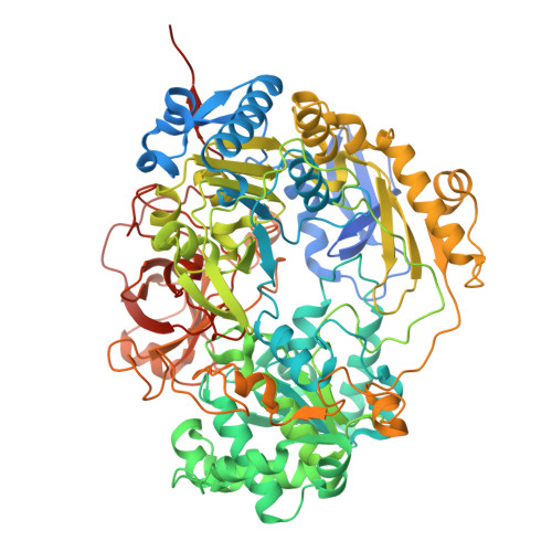
    9H4T

  • PubMed Abstract: 

    The enterobacterium Escherichia coli present in the human gut can reduce trimethylamine N-oxide (TMAO) to trimethylamine during anaerobic respiration. The TMAO reductase TorA is a monomeric, bis-molybdopterin guanine dinucleotide (bis-MGD) cofactor-containing enzyme that belongs to the dimethyl sulfoxide reductase family of molybdoenzymes. TorA is anchored to the membrane via TorC, a pentahemic c -type cytochrome which receives the electrons from the menaquinol pool. Here, we designed an expression system for the production of a stable soluble form of multiheme-containing TorC, providing, for the first time, the purification of a soluble pentahemic cytochrome- c from E. coli . Our focus was to investigate the interaction between TorA and soluble TorC to establish the electron transfer pathway. We solved the X-ray structure of E. coli TorA and performed chemical crosslinking of TorA and TorC. Another goal was to establish an activity assay that used the physiological electron transfer pathway instead of the commonly used unphysiological electron donors methylviologen or benzylviologen. An AlphaFold model including the crosslinking sites provided insights into the electron transfer between TorC C and the active site of TorA.


  • Organizational Affiliation
    • Department of Molecular Enzymology, Institute of Biochemistry and Biology, University of Potsdam, Karl-Liebknecht Str. 24-25, 14476 Potsdam, Germany.

Macromolecule Content 

  • Total Structure Weight: 195.8 kDa 
  • Atom Count: 14,495 
  • Modeled Residue Count: 1,585 
  • Deposited Residue Count: 1,696 
  • Unique protein chains: 1

Macromolecules

Find similar proteins by:|  3D Structure
Entity ID: 1
MoleculeChains  Sequence LengthOrganismDetailsImage
Trimethylamine-N-oxide reductase 1
A, B
848Escherichia coliMutation(s): 0 
Gene Names: torAb0997JW0982
EC: 1.7.2.3
UniProt
Find proteins for P33225 (Escherichia coli (strain K12))
Explore P33225 
Go to UniProtKB:  P33225
Entity Groups
Sequence Clusters30% Identity50% Identity70% Identity90% Identity95% Identity100% Identity
UniProt GroupP33225
Sequence Annotations
Expand
Reference Sequence

Small Molecules

Ligands 6 Unique
IDChains Name / Formula / InChI Key2D Diagram3D Interactions
PGD
(Subject of Investigation/LOI)

Query on PGD


Download:Ideal Coordinates CCD File
C [auth A],
D [auth A],
Y [auth B],
Z [auth B]
2-AMINO-5,6-DIMERCAPTO-7-METHYL-3,7,8A,9-TETRAHYDRO-8-OXA-1,3,9,10-TETRAAZA-ANTHRACEN-4-ONE GUANOSINE DINUCLEOTIDE
C20 H24 N10 O13 P2 S2
SOFMTHSWCZNJTQ-ILXWUORBSA-N
1PE

Query on 1PE


Download:Ideal Coordinates CCD File
DA [auth B]PENTAETHYLENE GLYCOL
C10 H22 O6
JLFNLZLINWHATN-UHFFFAOYSA-N
PGE

Query on PGE


Download:Ideal Coordinates CCD File
CA [auth B],
EA [auth B],
L [auth A],
M [auth A],
N [auth A]
TRIETHYLENE GLYCOL
C6 H14 O4
ZIBGPFATKBEMQZ-UHFFFAOYSA-N
MLT

Query on MLT


Download:Ideal Coordinates CCD File
FA [auth B]
GA [auth B]
HA [auth B]
O [auth A]
P [auth A]
FA [auth B],
GA [auth B],
HA [auth B],
O [auth A],
P [auth A],
Q [auth A],
R [auth A]
D-MALATE
C4 H6 O5
BJEPYKJPYRNKOW-UWTATZPHSA-N
PEG

Query on PEG


Download:Ideal Coordinates CCD File
BA [auth B]
F [auth A]
G [auth A]
H [auth A]
I [auth A]
BA [auth B],
F [auth A],
G [auth A],
H [auth A],
I [auth A],
IA [auth B],
J [auth A],
JA [auth B],
K [auth A],
S [auth A],
T [auth A],
U [auth A],
V [auth A],
W [auth A],
X [auth A]
DI(HYDROXYETHYL)ETHER
C4 H10 O3
MTHSVFCYNBDYFN-UHFFFAOYSA-N
6MO
(Subject of Investigation/LOI)

Query on 6MO


Download:Ideal Coordinates CCD File
AA [auth B],
E [auth A]
MOLYBDENUM(VI) ION
Mo
HCNGUXXTNNIKCQ-UHFFFAOYSA-N

Experimental Data & Validation

Experimental Data

  • Method: X-RAY DIFFRACTION
  • Resolution: 1.86 Å
  • R-Value Free:  0.289 (Depositor), 0.278 (DCC) 
  • R-Value Work:  0.235 (Depositor) 
  • R-Value Observed: 0.236 (Depositor) 
Space Group: P 1 21 1
Unit Cell:
Length ( Å )Angle ( ˚ )
a = 93.662α = 90
b = 117.467β = 103.25
c = 100.053γ = 90
Software Package:
Software NamePurpose
REFMACrefinement
autoXDSdata reduction
XSCALEdata scaling
PHASERphasing

Structure Validation

View Full Validation Report


Ligand Structure Quality Assessment 


Entry History 

& Funding Information

Deposition Data


Funding OrganizationLocationGrant Number
German Research Foundation (DFG)GermanyEXC 2008-390540038

Revision History  (Full details and data files)

  • Version 1.0: 2025-02-26
    Type: Initial release