


A novel copper-tyrosyl site among type-3 copper proteins
Englund, A.N.B., Rohr, A.K., Dalleywater, E.L.To be published.
Experimental Data Snapshot
Starting Model: experimental
View more details
wwPDB Validation 3D Report Full Report

Entity ID: 1 | |||||
|---|---|---|---|---|---|
| Molecule | Chains | Sequence Length | Organism | Details | Image |
| Tyrosinase | 303 | Priestia megaterium | Mutation(s): 1 | 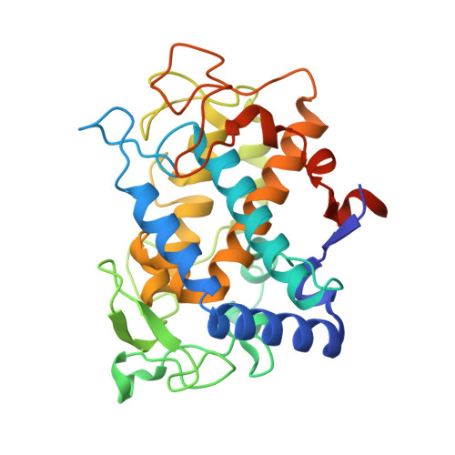 | |
UniProt | |||||
Entity Groups | |||||
| Sequence Clusters | 30% Identity50% Identity70% Identity90% Identity95% Identity100% Identity | ||||
| UniProt Group | B2ZB02 | ||||
Sequence AnnotationsExpand | |||||
Reference Sequence | |||||
| Ligands 1 Unique | |||||
|---|---|---|---|---|---|
| ID | Chains | Name / Formula / InChI Key | 2D Diagram | 3D Interactions | |
| CU (Subject of Investigation/LOI) Download:Ideal Coordinates CCD File | C [auth A], D [auth A], E [auth B], F [auth B] | COPPER (II) ION Cu JPVYNHNXODAKFH-UHFFFAOYSA-N |  | ||
| Length ( Å ) | Angle ( ˚ ) |
|---|---|
| a = 47.514 | α = 90 |
| b = 78.039 | β = 90 |
| c = 150.075 | γ = 90 |
| Software Name | Purpose |
|---|---|
| PHENIX | phasing |
| PHENIX | refinement |
| Funding Organization | Location | Grant Number |
|---|---|---|
| Norwegian Research Council | Norway | 301022 |


