


Crystal structure of human NUDT14 in complex with an inhibitor (MA-955-9)
Balikci, E., Feyerherm, C., Bradshaw, W., Apostolidou, M., Adcock, C., McGown, A., Spencer, J., Huber, K.To be published.
Experimental Data Snapshot
Starting Model: experimental
View more details
Entity ID: 1 | |||||
|---|---|---|---|---|---|
| Molecule | Chains | Sequence Length | Organism | Details | Image |
| Uridine diphosphate glucose pyrophosphatase NUDT14 | 223 | Homo sapiens | Mutation(s): 0 Gene Names: NUDT14, UGPP EC: 3.6.1.45 (PDB Primary Data), 3.6.1 (UniProt), 3.6.1.13 (UniProt) | 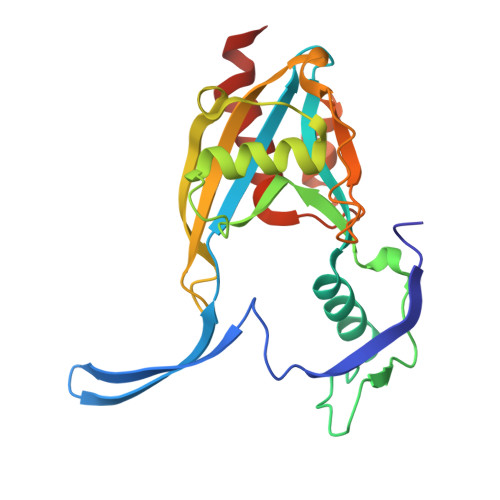 | |
UniProt & NIH Common Fund Data Resources | |||||
PHAROS: O95848 GTEx: ENSG00000183828 | |||||
Entity Groups | |||||
| Sequence Clusters | 30% Identity50% Identity70% Identity90% Identity95% Identity100% Identity | ||||
| UniProt Group | O95848 | ||||
Sequence AnnotationsExpand | |||||
Reference Sequence | |||||
| Ligands 6 Unique | |||||
|---|---|---|---|---|---|
| ID | Chains | Name / Formula / InChI Key | 2D Diagram | 3D Interactions | |
| A1IRF Download:Ideal Coordinates CCD File | C [auth A], H [auth A], L [auth B] | 1-(1-methylpiperidin-4-yl)-3-(3-phenoxyphenyl)pyrazolo[3,4-d]pyrimidin-4-amine C23 H24 N6 O JWZBPGIFSZRCBO-UHFFFAOYSA-N |  | ||
| PEG Download:Ideal Coordinates CCD File | F [auth A], G [auth A], M [auth B] | DI(HYDROXYETHYL)ETHER C4 H10 O3 MTHSVFCYNBDYFN-UHFFFAOYSA-N |  | ||
| GOL Download:Ideal Coordinates CCD File | D [auth A] | GLYCEROL C3 H8 O3 PEDCQBHIVMGVHV-UHFFFAOYSA-N |  | ||
| DMS Download:Ideal Coordinates CCD File | J [auth A], K [auth A], O [auth B] | DIMETHYL SULFOXIDE C2 H6 O S IAZDPXIOMUYVGZ-UHFFFAOYSA-N |  | ||
| ACY Download:Ideal Coordinates CCD File | E [auth A], N [auth B] | ACETIC ACID C2 H4 O2 QTBSBXVTEAMEQO-UHFFFAOYSA-N |  | ||
| MG Download:Ideal Coordinates CCD File | I [auth A] | MAGNESIUM ION Mg JLVVSXFLKOJNIY-UHFFFAOYSA-N |  | ||
| Length ( Å ) | Angle ( ˚ ) |
|---|---|
| a = 62.989 | α = 90 |
| b = 98.578 | β = 90 |
| c = 168.783 | γ = 90 |
| Software Name | Purpose |
|---|---|
| PHENIX | refinement |
| DIALS | data reduction |
| Aimless | data scaling |
| PHASER | phasing |
| Funding Organization | Location | Grant Number |
|---|---|---|
| European Union (EU) | European Union | 875510 |


