


A potent macrocyclic peptide modulator of Rab27a identified by RaPID screening
Petracca, R., Tersa, M., De Vita, E., Morgan, M.R., Cota, E., Tate, E.W.To be published.
Experimental Data Snapshot
Starting Model: experimental
View more details
Entity ID: 1 | |||||
|---|---|---|---|---|---|
| Molecule | Chains | Sequence Length | Organism | Details | Image |
| Ras-related protein Rab-27A | 184 | Homo sapiens | Mutation(s): 3 Gene Names: RAB27A, RAB27 EC: 3.6.5.2 |  | |
UniProt & NIH Common Fund Data Resources | |||||
PHAROS: P51159 GTEx: ENSG00000069974 | |||||
Entity Groups | |||||
| Sequence Clusters | 30% Identity50% Identity70% Identity90% Identity95% Identity100% Identity | ||||
| UniProt Group | P51159 | ||||
Sequence AnnotationsExpand | |||||
Reference Sequence | |||||
Entity ID: 2 | |||||
|---|---|---|---|---|---|
| Molecule | Chains | Sequence Length | Organism | Details | Image |
| Cyclic peptide IMP-2660 | C [auth D], D [auth F] | 14 | synthetic construct | Mutation(s): 0 | 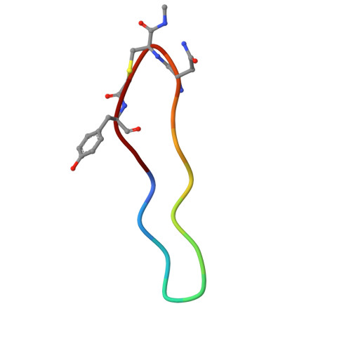 |
| Ligands 3 Unique | |||||
|---|---|---|---|---|---|
| ID | Chains | Name / Formula / InChI Key | 2D Diagram | 3D Interactions | |
| GNP Download:Ideal Coordinates CCD File | M [auth A], T [auth B] | PHOSPHOAMINOPHOSPHONIC ACID-GUANYLATE ESTER C10 H17 N6 O13 P3 UQABYHGXWYXDTK-UUOKFMHZSA-N |  | ||
| GOL Download:Ideal Coordinates CCD File | F [auth A] G [auth A] H [auth A] I [auth A] J [auth A] | GLYCEROL C3 H8 O3 PEDCQBHIVMGVHV-UHFFFAOYSA-N |  | ||
| MG Download:Ideal Coordinates CCD File | E [auth A], N [auth B] | MAGNESIUM ION Mg JLVVSXFLKOJNIY-UHFFFAOYSA-N |  | ||
| Length ( Å ) | Angle ( ˚ ) |
|---|---|
| a = 70.357 | α = 90 |
| b = 72.714 | β = 90 |
| c = 91.658 | γ = 90 |
| Software Name | Purpose |
|---|---|
| PHENIX | refinement |
| xia2 | data reduction |
| DIALS | data scaling |
| PHASER | phasing |
| Funding Organization | Location | Grant Number |
|---|---|---|
| Cancer Research UK | United Kingdom | C29637/A20781 |
| Cancer Research UK | United Kingdom | C29637/A26892 |
| H2020 Marie Curie Actions of the European Commission | European Union | 797995 |


