9GVA | pdb_00009gva

Crystal structure of the gamma carbonic anhydrase from Porphyromonas gingivalis


Experimental Data Snapshot

  • Method: X-RAY DIFFRACTION
  • Resolution: 2.40 Å
  • R-Value Free: 
    0.197 (Depositor), 0.199 (DCC) 
  • R-Value Work: 
    0.165 (Depositor), 0.175 (DCC) 

Starting Model: in silico
View more details

wwPDB Validation 3D Report Full Report

Validation slider image for 9GVA

This is version 1.0 of the entry. See complete history

Literature

Kinetic and structural studies of gamma-carbonic anhydrase from the oral pathogen Porphyromonas gingivalis.

Ferraroni, M.Angeli, A.De Luca, V.Capasso, C.Supuran, C.T.

(2024) J Struct Biol 217: 108154-108154

  • DOI: https://doi.org/10.1016/j.jsb.2024.108154
  • Primary Citation Related Structures: 
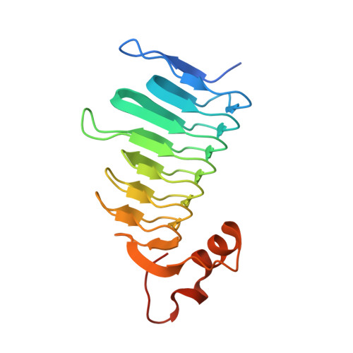
    9GVA

  • PubMed Abstract: 

    Porphyromonas gingivalis, a key pathogen in periodontal, plays a critical role in systemic pathologiesdiseases by evading host defence mechanisms and invading periodontal tissues. Targeting its virulence mechanisms and overcoming drug resistance are essential steps toward effective therapeutic development. In this study, we focused on the Carbonic Anhydrase (CA, EC: 4.2.1.1) encoded by P. gingivalis as a potential drug target. We determined the crystal structure of PgiCA γ at a resolution of 2.4 Å and conducted kinetic characterization. The structure revealed that active PgiCA γ forms a trimer, with each monomer comprising a left-handed β-helix capped by a C-terminal α-helix and coordinated to a catalytic zinc ion through three histidine residues. Interestingly, one monomer displayed an atypical α-helix conformation, likely due to close interactions with neighbouring trimers within the crystal lattice (a probable crystallographic artefact). These findings provide new insights into the structural and functional properties of PgiCA γ, emphasizing its potential as a target for the development of novel anti-virulence therapies against P. gingivalis.


  • Organizational Affiliation
    • Department of Chemistry "Ugo Schiff", University of Florence, Via Della Lastruccia 3-13, 50019, Sesto Fiorentino, Italy.

Macromolecule Content 

  • Total Structure Weight: 62.75 kDa 
  • Atom Count: 4,024 
  • Modeled Residue Count: 518 
  • Deposited Residue Count: 576 
  • Unique protein chains: 1

Macromolecules

Find similar proteins by:|  3D Structure
Entity ID: 1
MoleculeChains  Sequence LengthOrganismDetailsImage
Hexapeptide transferase family proteinA [auth AAA],
B [auth BBB],
C [auth CCC]
192Porphyromonas gingivalisMutation(s): 0 
Gene Names: PG_1211
UniProt
Find proteins for Q7MV79 (Porphyromonas gingivalis (strain ATCC BAA-308 / W83))
Explore Q7MV79 
Go to UniProtKB:  Q7MV79
Entity Groups
Sequence Clusters30% Identity50% Identity70% Identity90% Identity95% Identity100% Identity
UniProt GroupQ7MV79
Sequence Annotations
Expand
Reference Sequence

Experimental Data & Validation

Experimental Data

  • Method: X-RAY DIFFRACTION
  • Resolution: 2.40 Å
  • R-Value Free:  0.197 (Depositor), 0.199 (DCC) 
  • R-Value Work:  0.165 (Depositor), 0.175 (DCC) 
Space Group: P 65
Unit Cell:
Length ( Å )Angle ( ˚ )
a = 120.752α = 90
b = 120.752β = 90
c = 91.31γ = 120
Software Package:
Software NamePurpose
REFMACrefinement
XDSdata reduction
Aimlessdata scaling
REFMACphasing

Structure Validation

View Full Validation Report


Entry History 

& Funding Information

Deposition Data


Funding OrganizationLocationGrant Number
Not funded--

Revision History  (Full details and data files)

  • Version 1.0: 2024-12-25
    Type: Initial release