9GBR

Human Angiotensin-1 converting enzyme N-domain in complex with a diprolyl inhibitor- SG17


Experimental Data Snapshot

  • Method: X-RAY DIFFRACTION
  • Resolution: 1.90 Å
  • R-Value Free: 
    0.225 (Depositor), 0.227 (DCC) 
  • R-Value Work: 
    0.188 (Depositor), 0.188 (DCC) 

Starting Model: experimental
View more details

wwPDB Validation   3D Report Full Report


Ligand Structure Quality Assessment 

Created with Raphaël 2.3.0Worse 01 BetterLigand structure goodness of fit to experimental dataBest fitted A1IJXClick on this verticalbar to view details

This is version 1.0 of the entry. See complete history


Literature

Molecular basis of human angiotensin-1 converting enzyme inhibition by a series of diprolyl-derived compounds.

Gregory, K.S.Cozier, G.E.Fienberg, S.Chibale, K.Sturrock, E.D.Acharya, K.R.

(2025) FEBS J 

  • DOI: https://doi.org/10.1111/febs.17384
  • Primary Citation of Related Structures:  
    9GBL, 9GBM, 9GBN, 9GBO, 9GBP, 9GBQ, 9GBR, 9GBS

  • PubMed Abstract: 

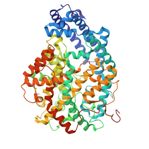
    Angiotensin-1-converting enzyme (ACE) is a zinc-dependent carboxypeptidase of therapeutic interest for the treatment of hypertension, inflammation and fibrosis. It consists of two homologous N and C catalytic domains, nACE and cACE, respectively. Unfortunately, the current clinically available ACE inhibitors produce undesirable side effects due to the nonselective inhibition of these domains. Through structure-based drug design, we previously identified a series of diprolyl-derived inhibitors (SG3, SG15, SG16, SG17 and SG18) in an attempt to specifically target nACE. Only one compound, SG16, possessed significant nACEselectivity. The previously determined 16-nACE crystal structure (nACE:SG16) suggested interactions with Tyr369 (Phe381 in cACE) are responsible for this selectivity. To better understand the molecular basis for the lack of selectivity in the remaining compounds, we have cocrystallised nACE in complex with SG3, SG15, SG17 and SG18 and cACE in complex with SG3, SG15, SG16 and SG18 and determined their structures at high resolution. Apart from the catalytic residues, these structures further highlight the importance of residues distal to the active site that may play an important role in the design of domain-selective inhibitors of ACE.


  • Organizational Affiliation

    Department of Life Sciences, University of Bath, UK.


Macromolecules
Find similar proteins by:  (by identity cutoff)  |  3D Structure
Entity ID: 1
MoleculeChains Sequence LengthOrganismDetailsImage
Angiotensin-converting enzyme, soluble form
A, B
628Homo sapiensMutation(s): 8 
Gene Names: ACEDCPDCP1
EC: 3.4.15.1
UniProt & NIH Common Fund Data Resources
Find proteins for P12821 (Homo sapiens)
Explore P12821 
Go to UniProtKB:  P12821
PHAROS:  P12821
GTEx:  ENSG00000159640 
Entity Groups  
Sequence Clusters30% Identity50% Identity70% Identity90% Identity95% Identity100% Identity
UniProt GroupP12821
Glycosylation
Glycosylation Sites: 3Go to GlyGen: P12821-1
Sequence Annotations
Expand
  • Reference Sequence
Oligosaccharides

Help

Entity ID: 2
MoleculeChains Length2D Diagram Glycosylation3D Interactions
2-acetamido-2-deoxy-beta-D-glucopyranose-(1-4)-2-acetamido-2-deoxy-beta-D-glucopyranose
C
2N-Glycosylation
Glycosylation Resources
GlyTouCan:  G42666HT
GlyCosmos:  G42666HT
GlyGen:  G42666HT
Entity ID: 3
MoleculeChains Length2D Diagram Glycosylation3D Interactions
alpha-L-fucopyranose-(1-6)-2-acetamido-2-deoxy-beta-D-glucopyranose
D
2N-Glycosylation
Glycosylation Resources
GlyTouCan:  G86851RC
GlyCosmos:  G86851RC
GlyGen:  G86851RC
Entity ID: 4
MoleculeChains Length2D Diagram Glycosylation3D Interactions
beta-D-mannopyranose-(1-4)-2-acetamido-2-deoxy-beta-D-glucopyranose-(1-4)-[alpha-L-fucopyranose-(1-6)]2-acetamido-2-deoxy-beta-D-glucopyranose
E
4N-Glycosylation
Glycosylation Resources
GlyTouCan:  G32152BH
GlyCosmos:  G32152BH
GlyGen:  G32152BH
Small Molecules
Ligands 7 Unique
IDChains Name / Formula / InChI Key2D Diagram3D Interactions
A1IJX (Subject of Investigation/LOI)
Query on A1IJX

Download Ideal Coordinates CCD File 
J [auth A],
P [auth B]
(2S)-1-[(2S)-2-[[(1S)-1-[(2S)-1-[(2S)-2-azanyl-5-oxidanyl-5-oxidanylidene-pentanoyl]pyrrolidin-2-yl]-2-oxidanyl-2-oxidanylidene-ethyl]amino]propanoyl]pyrrolidine-2-carboxylic acid
C19 H30 N4 O8
IGMKHUYAVWRMEJ-CXOVXGEYSA-N
P33
Query on P33

Download Ideal Coordinates CCD File 
L [auth B]3,6,9,12,15,18-HEXAOXAICOSANE-1,20-DIOL
C14 H30 O8
XPJRQAIZZQMSCM-UHFFFAOYSA-N
NAG
Query on NAG

Download Ideal Coordinates CCD File 
F [auth A],
G [auth A]
2-acetamido-2-deoxy-beta-D-glucopyranose
C8 H15 N O6
OVRNDRQMDRJTHS-FMDGEEDCSA-N
PG4
Query on PG4

Download Ideal Coordinates CCD File 
M [auth B]TETRAETHYLENE GLYCOL
C8 H18 O5
UWHCKJMYHZGTIT-UHFFFAOYSA-N
PGE
Query on PGE

Download Ideal Coordinates CCD File 
K [auth B]TRIETHYLENE GLYCOL
C6 H14 O4
ZIBGPFATKBEMQZ-UHFFFAOYSA-N
ZN
Query on ZN

Download Ideal Coordinates CCD File 
H [auth A],
N [auth B]
ZINC ION
Zn
PTFCDOFLOPIGGS-UHFFFAOYSA-N
CL
Query on CL

Download Ideal Coordinates CCD File 
I [auth A],
O [auth B]
CHLORIDE ION
Cl
VEXZGXHMUGYJMC-UHFFFAOYSA-M
Experimental Data & Validation

Experimental Data

  • Method: X-RAY DIFFRACTION
  • Resolution: 1.90 Å
  • R-Value Free:  0.225 (Depositor), 0.227 (DCC) 
  • R-Value Work:  0.188 (Depositor), 0.188 (DCC) 
Space Group: P 1
Unit Cell:
Length ( Å )Angle ( ˚ )
a = 73.053α = 88.38
b = 77.377β = 64.31
c = 82.914γ = 74.92
Software Package:
Software NamePurpose
REFMACrefinement
DIALSdata reduction
Aimlessdata scaling
PHASERphasing

Structure Validation

View Full Validation Report


Ligand Structure Quality Assessment 

Created with Raphaël 2.3.0Worse 01 BetterLigand structure goodness of fit to experimental dataBest fitted A1IJXClick on this verticalbar to view details

Entry History & Funding Information

Deposition Data


Funding OrganizationLocationGrant Number
Biotechnology and Biological Sciences Research Council (BBSRC)United KingdomBB/X001032/1

Revision History  (Full details and data files)

  • Version 1.0: 2025-01-22
    Type: Initial release