


Structure of Response regulator PleD in complex with c-diGMP and pppGpp
Dugelay, C., Jaboulay, C., Guzzo, M., Terradot, L.To be published.
Experimental Data Snapshot
Starting Model: experimental
View more details
Entity ID: 1 | |||||
|---|---|---|---|---|---|
| Molecule | Chains | Sequence Length | Organism | Details | Image |
| Response regulator PleD | 453 | Caulobacter vibrioides | Mutation(s): 0 Gene Names: pleD, CC_2462 EC: 2.7.7.65 | 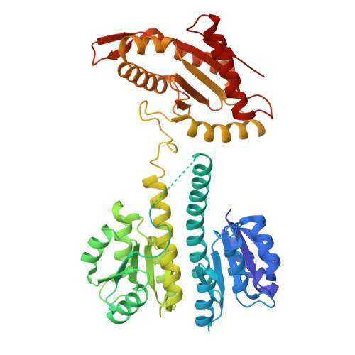 | |
UniProt | |||||
Entity Groups | |||||
| Sequence Clusters | 30% Identity50% Identity70% Identity90% Identity95% Identity100% Identity | ||||
| UniProt Group | Q9A5I5 | ||||
Sequence AnnotationsExpand | |||||
Reference Sequence | |||||
| Ligands 3 Unique | |||||
|---|---|---|---|---|---|
| ID | Chains | Name / Formula / InChI Key | 2D Diagram | 3D Interactions | |
| C2E (Subject of Investigation/LOI) Download:Ideal Coordinates CCD File | E [auth A] H [auth B] I [auth B] J [auth B] M [auth C] | 9,9'-[(2R,3R,3aS,5S,7aR,9R,10R,10aS,12S,14aR)-3,5,10,12-tetrahydroxy-5,12-dioxidooctahydro-2H,7H-difuro[3,2-d:3',2'-j][1,3,7,9,2,8]tetraoxadiphosphacyclododecine-2,9-diyl]bis(2-amino-1,9-dihydro-6H-purin-6-one) C20 H24 N10 O14 P2 PKFDLKSEZWEFGL-MHARETSRSA-N |  | ||
| 0O2 (Subject of Investigation/LOI) Download:Ideal Coordinates CCD File | F [auth A], K [auth B], N [auth C], Q [auth D] | guanosine 5'-(tetrahydrogen triphosphate) 3'-(trihydrogen diphosphate) C10 H18 N5 O20 P5 KCPMACXZAITQAX-UUOKFMHZSA-N |  | ||
| MG (Subject of Investigation/LOI) Download:Ideal Coordinates CCD File | G [auth A], L [auth B], O [auth C], T [auth D] | MAGNESIUM ION Mg JLVVSXFLKOJNIY-UHFFFAOYSA-N |  | ||
| Length ( Å ) | Angle ( ˚ ) |
|---|---|
| a = 132.571 | α = 90 |
| b = 132.571 | β = 90 |
| c = 508.411 | γ = 120 |
| Software Name | Purpose |
|---|---|
| PHENIX | refinement |
| Aimless | data scaling |
| XDS | data reduction |
| PHASER | phasing |
| Funding Organization | Location | Grant Number |
|---|---|---|
| ATIP-Avenir | France | -- |


