9G7H | pdb_00009g7h

Human Sirt6 in complex with ADP-ribose and the inhibitor 2-Pr


Experimental Data Snapshot

  • Method: X-RAY DIFFRACTION
  • Resolution: 1.75 Å
  • R-Value Free: 
    0.202 (Depositor), 0.205 (DCC) 
  • R-Value Work: 
    0.167 (Depositor), 0.172 (DCC) 
  • R-Value Observed: 
    0.168 (Depositor) 

Starting Model: experimental
View more details

wwPDB Validation 3D Report Full Report

Validation slider image for 9G7H

Ligand Structure Quality Assessment 


This is version 1.1 of the entry. See complete history

Literature

Elucidating the Unconventional Binding Mode of a DNA-Encoded Library Hit Provides a Blueprint for Sirtuin 6 Inhibitor Development.

You, W.Montoya, A.L.Dana, S.Franzini, R.M.Steegborn, C.

(2024) ChemMedChem 19: e202400273-e202400273

  • DOI: https://doi.org/10.1002/cmdc.202400273
  • Primary Citation Related Structures: 
    9G7H

  • PubMed Abstract: 

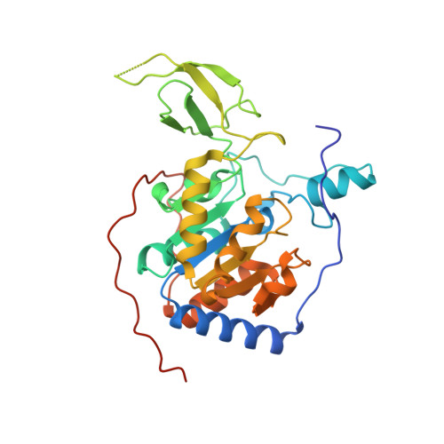
    Sirtuin 6 (Sirt6), an NAD+-dependent deacylase, has emerged as a promising target for aging-related diseases and cancer. Advancing the medicinal chemistry of Sirt6 modulators is crucial for the development of chemical probes aimed at unraveling the intricate biological functions of Sirt6 and unlocking its therapeutic potential. A proprietary DNA-encoded library yielded Sirt6 inhibitor 2-Pr, displaying remarkable inhibitory activity and isoform-selectivity, and featuring a chemical structure distinct from reported Sirt6 modulators. In this study, we explore the inhibitory mechanism of 2-Pr, evaluating the impact of chemical modifications and presenting a crystal structure of the Sirt6/ADP-ribose/2-Pr complex. Notably, co-crystal structure analysis reveals an unexpected and unprecedented binding mode of Sirt6, with 2-Pr spanning the acyl channel of the enzyme, extending into the acetyl-lysine binding pocket, and reaching toward the C-site. This unique binding mode guides potential avenues for developing potent and selective Sirt6 inhibitors.


  • Organizational Affiliation
    • Bayreuth University, Biochemistry, GERMANY.

Macromolecule Content 

  • Total Structure Weight: 69.73 kDa 
  • Atom Count: 4,656 
  • Modeled Residue Count: 559 
  • Deposited Residue Count: 604 
  • Unique protein chains: 1

Macromolecules

Find similar proteins by:|  3D Structure
Entity ID: 1
MoleculeChains  Sequence LengthOrganismDetailsImage
NAD-dependent protein deacetylase sirtuin-6
A, B
302Homo sapiensMutation(s): 0 
Gene Names: SIRT6SIR2L6
EC: 2.3.1.286 (PDB Primary Data), 2.4.2 (UniProt), 2.3.1 (UniProt)
UniProt & NIH Common Fund Data Resources
Find proteins for Q8N6T7 (Homo sapiens)
Explore Q8N6T7 
Go to UniProtKB:  Q8N6T7
PHAROS:  Q8N6T7
GTEx:  ENSG00000077463 
Entity Groups
Sequence Clusters30% Identity50% Identity70% Identity90% Identity95% Identity100% Identity
UniProt GroupQ8N6T7
Sequence Annotations
Expand
Reference Sequence

Small Molecules

Ligands 4 Unique
IDChains Name / Formula / InChI Key2D Diagram3D Interactions
AR6

Query on AR6


Download:Ideal Coordinates CCD File
C [auth A],
K [auth B]
[(2R,3S,4R,5R)-5-(6-AMINOPURIN-9-YL)-3,4-DIHYDROXY-OXOLAN-2-YL]METHYL[HYDROXY-[[(2R,3S,4R,5S)-3,4,5-TRIHYDROXYOXOLAN-2-YL]METHOXY]PHOSPHORYL] HYDROGEN PHOSPHATE
C15 H23 N5 O14 P2
SRNWOUGRCWSEMX-ZQSHOCFMSA-N
A1IIZ
(Subject of Investigation/LOI)

Query on A1IIZ


Download:Ideal Coordinates CCD File
E [auth A]2,4-bis(oxidanylidene)-1-[2-oxidanylidene-2-[[(2S)-3-oxidanylidene-3-(propylamino)-2-[3-(3-pyridin-3-yl-1,2,4-oxadiazol-5-yl)propanoylamino]propyl]amino]ethyl]pyrimidine-5-carboxamide
C23 H27 N9 O7
WGTNFHHLZQGSEM-HNNXBMFYSA-N
SO4

Query on SO4


Download:Ideal Coordinates CCD File
F [auth A]
G [auth A]
H [auth A]
I [auth A]
J [auth A]
F [auth A],
G [auth A],
H [auth A],
I [auth A],
J [auth A],
M [auth B],
N [auth B]
SULFATE ION
O4 S
QAOWNCQODCNURD-UHFFFAOYSA-L
ZN

Query on ZN


Download:Ideal Coordinates CCD File
D [auth A],
L [auth B]
ZINC ION
Zn
PTFCDOFLOPIGGS-UHFFFAOYSA-N

Experimental Data & Validation

Experimental Data

  • Method: X-RAY DIFFRACTION
  • Resolution: 1.75 Å
  • R-Value Free:  0.202 (Depositor), 0.205 (DCC) 
  • R-Value Work:  0.167 (Depositor), 0.172 (DCC) 
  • R-Value Observed: 0.168 (Depositor) 
Space Group: P 63
Unit Cell:
Length ( Å )Angle ( ˚ )
a = 91.466α = 90
b = 91.466β = 90
c = 144.608γ = 120
Software Package:
Software NamePurpose
REFMACrefinement
XDSdata scaling
XDSdata reduction
PHASERphasing

Structure Validation

View Full Validation Report


Ligand Structure Quality Assessment 


Entry History 

& Funding Information

Deposition Data


Funding OrganizationLocationGrant Number
Not funded--

Revision History  (Full details and data files)

  • Version 1.0: 2024-08-14
    Type: Initial release
  • Version 1.1: 2024-10-30
    Changes: Database references, Structure summary