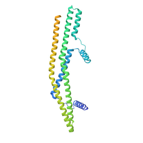
Peripheral membrane protein endophilin B1 probes, perturbs and permeabilizes lipid bilayers.
Thorlacius, A., Rulev, M., Sundberg, O., Sundborger-Lunna, A.(2025) Commun Biol 8: 182-182
- PubMed: 39910321
- DOI: https://doi.org/10.1038/s42003-025-07610-1
- Primary Citation of Related Structures:
9G2R, 9G2U, 9G2W - PubMed Abstract:
Bin/Amphiphysin/Rvs167 (BAR) domain containing proteins are peripheral membrane proteins that regulate intracellular membrane curvature. BAR protein endophilin B1 plays a key role in multiple cellular processes critical for oncogenesis, including autophagy and apoptosis. Amphipathic regions in endophilin B1 drive membrane association and tubulation through membrane scaffolding. Our understanding of exactly how BAR proteins like endophilin B1 promote highly diverse intracellular membrane remodeling events in the cell is severely limited due to lack of high-resolution structural information. Here we present the highest resolution cryo-EM structure of a BAR protein to date and the first structures of a BAR protein bound to a lipid bicelle. Using neural networks, we can effectively sort particle species of different stoichiometries, revealing the tremendous flexibility of post-membrane binding, pre-polymer BAR dimer organization and membrane deformation. We also show that endophilin B1 efficiently permeabilizes negatively charged liposomes that contain mitochondria-specific lipid cardiolipin and propose a new model for Bax-mediated cell death.
- Department of Cell and Molecular Biology, Uppsala University, Uppsala, Sweden.
Organizational Affiliation:


