9G0J | pdb_00009g0j

Structure of the PRO-PRO endopeptidase (PPEP-3) from Geobacillus thermodenitrificans


Experimental Data Snapshot

  • Method: X-RAY DIFFRACTION
  • Resolution: 1.55 Å
  • R-Value Free: 
    0.198 (Depositor), 0.199 (DCC) 
  • R-Value Work: 
    0.173 (Depositor), 0.174 (DCC) 
  • R-Value Observed: 
    0.174 (Depositor) 

Starting Model: experimental
View more details

wwPDB Validation 3D Report Full Report

Validation slider image for 9G0J

Ligand Structure Quality Assessment 


This is version 1.2 of the entry. See complete history

Literature

Structural analyses and substrate profiling of PPEP-3 provide new insights into the molecular basis of Pro-Pro endopeptidase specificity.

Claushuis, B.Wojtalla, F.Papenhagen, L.Cordfunke, R.A.de Ru, A.H.van Leeuwen, H.C.Corver, J.Hensbergen, P.J.Baumann, U.

(2026) iScience 29: 114360-114360

  • DOI: https://doi.org/10.1016/j.isci.2025.114360
  • Primary Citation Related Structures: 
    9G0J, 9G3T, 9G5J

  • PubMed Abstract: 

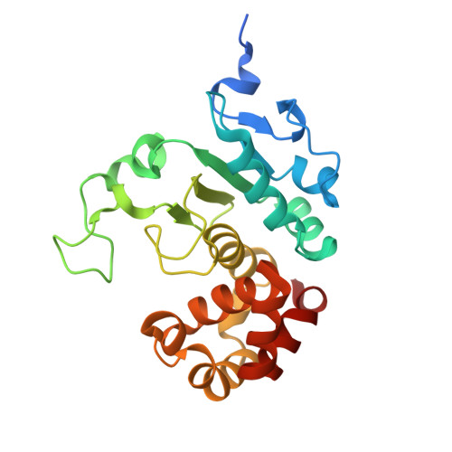
    Pro-Pro endopeptidases (PPEPs) are secreted bacterial enzymes that uniquely cleave peptide bonds between adjacent proline residues. Their active site accommodates six substrate residues (P3 to P3'), with interactions at these positions determining specificity. In this study, we investigated the substrate specificity of PPEP-3 from Geobacillus thermodenitrificans using synthetic peptide libraries and liquid chromatography-tandem mass spectrometry (LC-MS/MS). We also determined the atomic structures of PPEP-3 in unbound and substrate-bound forms. By correlating substrate profiling with structural data, we identified key mechanisms influencing PPEP-3 specificity. This integrated analysis reveals stark differences in specificity for the P2 and P2' positions compared to other PPEPs, most notably Tyr161 and Phe191, which shape the substrate-binding cleft and influence the accommodation of side chains at these positions. Combining comprehensive substrate profiling with structural analyses offers a powerful approach to uncover the molecular basis of protease function.


  • Organizational Affiliation
    • Center for Proteomics and Metabolomics, Leiden University Medical Center, Leiden 2333 ZA, the Netherlands.

Macromolecule Content 

  • Total Structure Weight: 53.6 kDa 
  • Atom Count: 3,731 
  • Modeled Residue Count: 421 
  • Deposited Residue Count: 460 
  • Unique protein chains: 1

Macromolecules

Find similar proteins by:|  3D Structure
Entity ID: 1
MoleculeChains  Sequence LengthOrganismDetailsImage
ATLF-like domain-containing protein
A, B
230Geobacillus thermodenitrificans NG80-2Mutation(s): 0 
Gene Names: GTNG_1672
UniProt
Find proteins for A4INY2 (Geobacillus thermodenitrificans (strain NG80-2))
Explore A4INY2 
Go to UniProtKB:  A4INY2
Entity Groups
Sequence Clusters30% Identity50% Identity70% Identity90% Identity95% Identity100% Identity
UniProt GroupA4INY2
Sequence Annotations
Expand
Reference Sequence

Small Molecules

Ligands 4 Unique
IDChains Name / Formula / InChI Key2D Diagram3D Interactions
BCN
(Subject of Investigation/LOI)

Query on BCN


Download:Ideal Coordinates CCD File
D [auth A],
H [auth B]
BICINE
C6 H13 N O4
FSVCELGFZIQNCK-UHFFFAOYSA-N
PEG

Query on PEG


Download:Ideal Coordinates CCD File
E [auth A],
I [auth B],
J [auth B]
DI(HYDROXYETHYL)ETHER
C4 H10 O3
MTHSVFCYNBDYFN-UHFFFAOYSA-N
ZN
(Subject of Investigation/LOI)

Query on ZN


Download:Ideal Coordinates CCD File
C [auth A],
G [auth B]
ZINC ION
Zn
PTFCDOFLOPIGGS-UHFFFAOYSA-N
EDO

Query on EDO


Download:Ideal Coordinates CCD File
F [auth A]1,2-ETHANEDIOL
C2 H6 O2
LYCAIKOWRPUZTN-UHFFFAOYSA-N

Experimental Data & Validation

Experimental Data

  • Method: X-RAY DIFFRACTION
  • Resolution: 1.55 Å
  • R-Value Free:  0.198 (Depositor), 0.199 (DCC) 
  • R-Value Work:  0.173 (Depositor), 0.174 (DCC) 
  • R-Value Observed: 0.174 (Depositor) 
Space Group: P 41 21 2
Unit Cell:
Length ( Å )Angle ( ˚ )
a = 126.247α = 90
b = 126.247β = 90
c = 64.043γ = 90
Software Package:
Software NamePurpose
PHENIXrefinement
XSCALEdata scaling
XDSdata reduction
PHASERphasing

Structure Validation

View Full Validation Report


Ligand Structure Quality Assessment 


Entry History 

& Funding Information

Deposition Data


Funding OrganizationLocationGrant Number
Netherlands Organisation for Scientific Research (NWO)NetherlandsOCENW.KLEIN.103

Revision History  (Full details and data files)

  • Version 1.0: 2025-07-16
    Type: Initial release
  • Version 1.1: 2026-02-04
    Changes: Data collection, Database references
  • Version 1.2: 2026-02-18
    Changes: Database references