Sulfated mannan of diatoms selects host-specific microbiota in the sunlit ocean.
Krull, J., Sidhu, C., Solanki, V., Bligh, M., Rossler, L., Singh, R.K., Huang, G., Robb, C.S., Teeling, H., Seeberger, P.H., Schweder, T., Crawford, C.J., Hehemann, J.H.(2026) Microbiome 14
- PubMed: 41888912 Search on PubMedSearch on PubMed Central
- DOI: https://doi.org/10.1186/s40168-026-02379-9
- Primary Citation Related Structures:
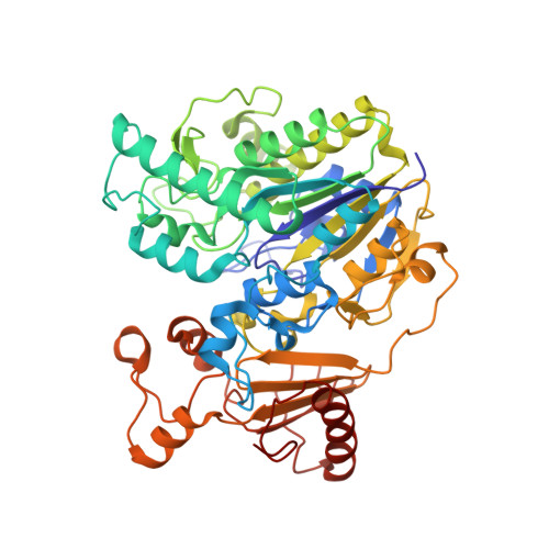
9FVT - PubMed Abstract:
Diatoms, a keystone phylum in Earth's ecosystems, are responsible for substantial oxygen production and the fixation of carbon dioxide in the form of carbohydrates that fuel global food webs. They host diverse prokaryotes, yet how diatoms preferentially recruit those with complementary metabolic traits remains unknown. We discovered that diatoms exude a C6-sulfated α-1,3-mannan that serves as a selective carbon source for adapted Polaribacter. Its structure was resolved using NMR spectroscopy, chromatography, chemical synthesis, and enzymatic dissection. Biochemical, physiological, and structural analyses demonstrated, that specialized Bacteroidota employ a four-enzyme pathway to metabolize this glycan. Metagenomic and transcriptomic data revealed that sulfated mannan utilization loci are globally abundant and actively expressed in surface ocean bacterioplankton. Because this mannan provides only carbon, oxygen, sulfur, and hydrogen, bacteria must obtain other essential elements elsewhere, reinforcing metabolic interdependence. Together, these results define a chemically specific interaction between diatoms and specialized bacteria that is mediated by a single sulfated polysaccharide and a dedicated four-enzyme degradation pathway. Presence of this pathway in marine metagenomes and transcriptomes indicates that a sulfated mannan from diatoms exerts selection pressure in the sunlit ocean microbiome. Video Abstract.
- Faculty of Chemistry & Biology, BIOM, University of Bremen, Bremen, Germany.
Organizational Affiliation: