9FSW | pdb_00009fsw

ClpP from Staphylococcus epidermidis with glycerol in some of the catalytic sites.


Experimental Data Snapshot

  • Method: X-RAY DIFFRACTION
  • Resolution: 2.39 Å
  • R-Value Free: 
    0.233 (Depositor), 0.231 (DCC) 
  • R-Value Work: 
    0.199 (Depositor), 0.199 (DCC) 
  • R-Value Observed: 
    0.201 (Depositor) 

Starting Model: experimental
View more details

wwPDB Validation 3D Report Full Report

Validation slider image for 9FSW

This is version 1.0 of the entry. See complete history

Literature

Molecular insights into the dynamic modulation of bacterial ClpP function and oligomerization by peptidomimetic boronate compounds.

Alves Franca, B.Falke, S.Rohde, H.Betzel, C.

(2024) Sci Rep 14: 2572

  • DOI: https://doi.org/10.1038/s41598-024-51787-0
  • Primary Citation Related Structures: 
    8CJ4, 8QYF, 9FSW, 9G6I, 9G72

  • PubMed Abstract: 

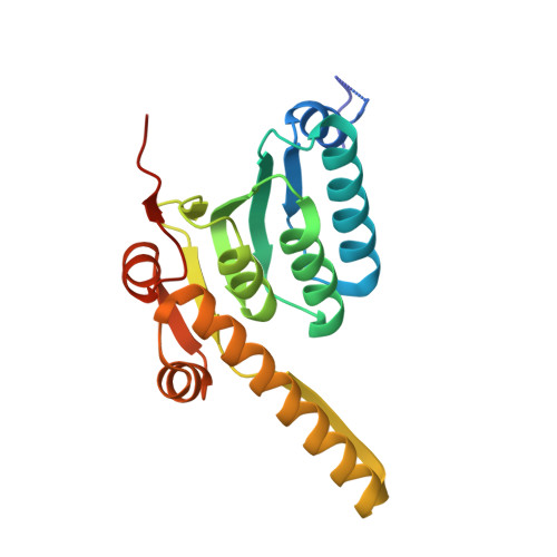
    Bacterial caseinolytic protease P subunit (ClpP) is important and vital for cell survival and infectivity. Recent publications describe and discuss the complex structure-function relationship of ClpP and its processive activity mediated by 14 catalytic sites. Even so, there are several aspects yet to be further elucidated, such as the paradoxical allosteric modulation of ClpP by peptidomimetic boronates. These compounds bind to all catalytic sites, and in specific conditions, they stimulate a dysregulated degradation of peptides and globular proteins, instead of inhibiting the enzymatic activity, as expected for serine proteases in general. Aiming to explore and explain this paradoxical effect, we solved and refined the crystal structure of native ClpP from Staphylococcus epidermidis (Se), an opportunistic pathogen involved in nosocomial infections, as well as ClpP in complex with ixazomib at 1.90 Å and 2.33 Å resolution, respectively. The interpretation of the crystal structures, in combination with complementary biochemical and biophysical data, shed light on how ixazomib affects the ClpP conformational state and activity. Moreover, SEC-SAXS and DLS measurements show, for the first time, that a peptidomimetic boronate compound also induces the assembly of the tetradecameric structure from isolated homomeric heptameric rings of a gram-positive organism.


  • Organizational Affiliation
    • Institute of Biochemistry and Molecular Biology, Laboratory for Structural Biology of Infection and Inflammation, University of Hamburg, c/o DESY, Build. 22a, Notkestraße 85, 22607, Hamburg, Germany.

Macromolecule Content 

  • Total Structure Weight: 312.06 kDa 
  • Atom Count: 20,061 
  • Modeled Residue Count: 2,520 
  • Deposited Residue Count: 2,786 
  • Unique protein chains: 1

Macromolecules

Find similar proteins by:|  3D Structure
Entity ID: 1
MoleculeChains  Sequence LengthOrganismDetailsImage
ATP-dependent Clp protease proteolytic subunit
A, B, C, D, E
A, B, C, D, E, F, G, H, I, J, K, L, M, N
199Staphylococcus epidermidisMutation(s): 0 
Gene Names: 
EC: 3.4.21.92
UniProt
Find proteins for Q2G036 (Staphylococcus aureus (strain NCTC 8325 / PS 47))
Explore Q2G036 
Go to UniProtKB:  Q2G036
Entity Groups
Sequence Clusters30% Identity50% Identity70% Identity90% Identity95% Identity100% Identity
UniProt GroupQ2G036
Sequence Annotations
Expand
Reference Sequence

Small Molecules

Ligands 2 Unique
IDChains Name / Formula / InChI Key2D Diagram3D Interactions
MPD

Query on MPD


Download:Ideal Coordinates CCD File
AA [auth G]
CA [auth H]
EA [auth I]
FA [auth J]
GA [auth K]
AA [auth G],
CA [auth H],
EA [auth I],
FA [auth J],
GA [auth K],
HA [auth L],
IA [auth M],
KA [auth N],
O [auth A],
R [auth B],
S [auth C],
U [auth D],
W [auth E],
Y [auth F]
(4S)-2-METHYL-2,4-PENTANEDIOL
C6 H14 O2
SVTBMSDMJJWYQN-YFKPBYRVSA-N
GOL

Query on GOL


Download:Ideal Coordinates CCD File
BA [auth H]
DA [auth I]
JA [auth N]
LA [auth N]
P [auth B]
BA [auth H],
DA [auth I],
JA [auth N],
LA [auth N],
P [auth B],
Q [auth B],
T [auth D],
V [auth E],
X [auth F],
Z [auth G]
GLYCEROL
C3 H8 O3
PEDCQBHIVMGVHV-UHFFFAOYSA-N

Experimental Data & Validation

Experimental Data

  • Method: X-RAY DIFFRACTION
  • Resolution: 2.39 Å
  • R-Value Free:  0.233 (Depositor), 0.231 (DCC) 
  • R-Value Work:  0.199 (Depositor), 0.199 (DCC) 
  • R-Value Observed: 0.201 (Depositor) 
Space Group: P 1 21 1
Unit Cell:
Length ( Å )Angle ( ˚ )
a = 93.606α = 90
b = 123.989β = 91.184
c = 126.688γ = 90
Software Package:
Software NamePurpose
PHENIXrefinement
Aimlessdata scaling
XDSdata reduction
PHENIXphasing

Structure Validation

View Full Validation Report


Entry History 

& Funding Information

Deposition Data


Funding OrganizationLocationGrant Number
German Research Foundation (DFG)Germany3907159
German Federal Ministry for Education and ResearchGermany05K19GU4
German Federal Ministry for Education and ResearchGermany05K20GUB

Revision History  (Full details and data files)

  • Version 1.0: 2025-07-02
    Type: Initial release