


X-ray structure of 25-hydroxy steroid kinase (25-HSK)
Jacoby, C., Demmer, U., Warkentin, E., Ermler, U., Boll, M.To be published.
Experimental Data Snapshot
Entity ID: 1 | |||||
|---|---|---|---|---|---|
| Molecule | Chains | Sequence Length | Organism | Details | Image |
| Aminoglycoside phosphotransferase-like protein | 379 | Sterolibacterium denitrificans | Mutation(s): 0 Gene Names: SDENCHOL_21286 | 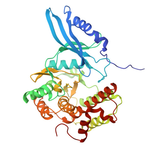 | |
UniProt | |||||
Find proteins for A0A7Z7HU23 (Sterolibacterium denitrificans) Explore A0A7Z7HU23 Go to UniProtKB: A0A7Z7HU23 | |||||
Entity Groups | |||||
| Sequence Clusters | 30% Identity50% Identity70% Identity90% Identity95% Identity100% Identity | ||||
| UniProt Group | A0A7Z7HU23 | ||||
Sequence AnnotationsExpand | |||||
Reference Sequence | |||||
| Ligands 4 Unique | |||||
|---|---|---|---|---|---|
| ID | Chains | Name / Formula / InChI Key | 2D Diagram | 3D Interactions | |
| A1IF2 (Subject of Investigation/LOI) Download:Ideal Coordinates CCD File | J [auth B], K [auth B] | 25-OH-cholest-1,4-diene-3-one C27 H42 O2 PFXYJIRDMVQBGX-REEZCCHISA-N |  | ||
| GOL Download:Ideal Coordinates CCD File | C [auth A] | GLYCEROL C3 H8 O3 PEDCQBHIVMGVHV-UHFFFAOYSA-N |  | ||
| ZN Download:Ideal Coordinates CCD File | E [auth A] F [auth A] G [auth A] H [auth A] I [auth A] | ZINC ION Zn PTFCDOFLOPIGGS-UHFFFAOYSA-N |  | ||
| MG Download:Ideal Coordinates CCD File | D [auth A], L [auth B] | MAGNESIUM ION Mg JLVVSXFLKOJNIY-UHFFFAOYSA-N |  | ||
| Length ( Å ) | Angle ( ˚ ) |
|---|---|
| a = 54.13 | α = 90 |
| b = 100.47 | β = 90 |
| c = 140.27 | γ = 90 |
| Software Name | Purpose |
|---|---|
| PHENIX | refinement |
| XSCALE | data scaling |
| XDS | data reduction |
| PHASER | phasing |
| Funding Organization | Location | Grant Number |
|---|---|---|
| Not funded | -- |


