SymProFold: Structural prediction of symmetrical biological assemblies.
Buhlheller, C., Sagmeister, T., Grininger, C., Gubensak, N., Sleytr, U.B., Uson, I., Pavkov-Keller, T.(2024) Nat Commun 15: 8152-8152
- PubMed: 39294115 Search on PubMedSearch on PubMed Central
- DOI: https://doi.org/10.1038/s41467-024-52138-3
- Primary Citation Related Structures:
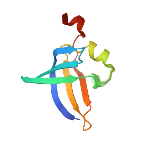
9FS9, 9FSA - PubMed Abstract:
Symmetry in nature often emerges from self-assembly processes and serves a wide range of functions. Cell surface layers (S-layers) form symmetrical lattices on many bacterial and archaeal cells, playing essential roles such as facilitating cell adhesion, evading the immune system, and protecting against environmental stress. However, the experimental structural characterization of these S-layers is challenging due to their self-assembly properties and high sequence variability. In this study, we introduce the SymProFold pipeline, which utilizes the high accuracy of AlphaFold-Multimer predictions to derive symmetrical assemblies from protein sequences, specifically focusing on two-dimensional S-layer arrays and spherical viral capsids. The pipeline tests all known symmetry operations observed in these systems (p1, p2, p3, p4, and p6) and identifies the most likely symmetry for the assembly. The predicted models were validated using available experimental data at the cellular level, and additional crystal structures were obtained to confirm the symmetry and interfaces of several SymProFold assemblies. Overall, the SymProFold pipeline enables the determination of symmetric protein assemblies linked to critical functions, thereby opening possibilities for exploring functionalities and designing targeted applications in diverse fields such as nanotechnology, biotechnology, medicine, and materials and environmental sciences.
- Institute of Molecular Biosciences, University of Graz, Graz, Austria.
Organizational Affiliation: