9FN3 | pdb_00009fn3

Crystal structure of the alkyltransferase ribozyme SAMURI co-crystalized with ProSeDMA


Experimental Data Snapshot

  • Method: X-RAY DIFFRACTION
  • Resolution: 2.90 Å
  • R-Value Free: 
    0.224 (Depositor), 0.237 (DCC) 
  • R-Value Work: 
    0.201 (Depositor), 0.211 (DCC) 
  • R-Value Observed: 
    0.202 (Depositor) 

wwPDB Validation 3D Report Full Report

Validation slider image for 9FN3

Ligand Structure Quality Assessment 


This is version 2.0 of the entry. See complete history

Literature

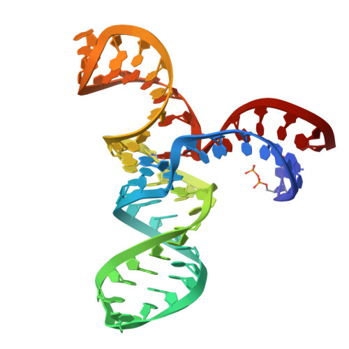
Structure and catalytic activity of the SAM-utilizing ribozyme SAMURI.

Chen, H.A.Okuda, T.Lenz, A.K.Scheitl, C.P.M.Schindelin, H.Hobartner, C.

(2025) Nat Chem Biol 

  • DOI: https://doi.org/10.1038/s41589-024-01808-w
  • Primary Citation Related Structures: 
    9FN2, 9FN3

  • PubMed Abstract: 

    Ribozymes that catalyze site-specific RNA modification have recently gained increasing interest for their ability to mimic methyltransferase enzymes and for their application to install molecular tags. Recently, we reported SAMURI as a site-specific alkyltransferase ribozyme using S-adenosylmethionine (SAM) or a stabilized analog to transfer a methyl or propargyl group to N 3 of an adenosine. Here, we report the crystal structures of SAMURI in the postcatalytic state. The structures reveal a three-helix junction with the catalytic core folded into four stacked layers, harboring the cofactor and the modified nucleotide. Detailed structure-activity analyses explain the cofactor scope and the structural basis for site selectivity. A structural comparison of SAMURI with SAM riboswitches sheds light on how the synthetic ribozyme overcomes the strategies of natural riboswitches to avoid self-methylation. Our results suggest that SAM and its analogs may serve as substrates for various RNA-catalyzed reactions, for which the corresponding ribozymes remain to be identified.


  • Organizational Affiliation
    • Institute of Organic Chemistry, Julius-Maximilians-Universität Würzburg, Würzburg, Germany.

Macromolecule Content 

  • Total Structure Weight: 38.68 kDa 
  • Atom Count: 2,590 
  • Modeled Residue Count: 116 
  • Deposited Residue Count: 116 
  • Unique nucleic acid chains: 1

Macromolecules

Find similar nucleic acids by:  (by identity cutoff) 
Entity ID: 1
MoleculeChains LengthOrganismImage
SAMURI-ProSeDMA
A, B
58synthetic construct
Sequence Annotations
Expand
Reference Sequence

Experimental Data & Validation

Experimental Data

  • Method: X-RAY DIFFRACTION
  • Resolution: 2.90 Å
  • R-Value Free:  0.224 (Depositor), 0.237 (DCC) 
  • R-Value Work:  0.201 (Depositor), 0.211 (DCC) 
  • R-Value Observed: 0.202 (Depositor) 
Space Group: P 42
Unit Cell:
Length ( Å )Angle ( ˚ )
a = 75.444α = 90
b = 75.444β = 90
c = 72.251γ = 90
Software Package:
Software NamePurpose
PHENIXrefinement
XDSdata scaling
XDSdata reduction
AutoSolphasing
Cootmodel building

Structure Validation

View Full Validation Report


Ligand Structure Quality Assessment 


Entry History 

& Funding Information

Deposition Data


Funding OrganizationLocationGrant Number
German Research Foundation (DFG)Germany--
European Research Council (ERC)European Union--

Revision History  (Full details and data files)

  • Version 1.0: 2025-02-05
    Type: Initial release
  • Version 2.0: 2025-02-12
    Type: Coordinate replacement
    Reason: Ligand geometry
    Changes: Advisory, Atomic model, Author supporting evidence, Data collection, Database references, Derived calculations, Refinement description, Structure summary